
Oncology and hematology

This collection groups existing oncology and haematology content across tumour types and care stages.
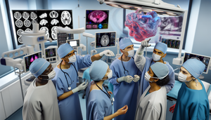
Modern operating room featuring a team of Hispanic surgeons utilizing a 3D holographic display to visualize tumors, emphasizing advanced oncological AR technology for margin delineation and surgical diagnosis.

Oncological Augmented Reality: 3D Tumor Visualization for Enhanced Margin Delineation and Surgical Diagnosis

Oncological augmented reality enhances surgical precision by visualizing tumors in 3D and delineating margins.

Read more

Hispanic scientist in a modern precision oncology laboratory, handling a liquid biopsy sample next to a genomic sequencing machine. The computer screen displays a detailed tumor DNA analysis, emphasizing advancements in advanced oncological diagnosis and precision genomics.

Liquid Biopsy and Precision Genomics: Advancing Oncological Diagnosis through Tumor DNA Analysis

Liquid biopsy and precision genomics are revolutionizing oncological diagnosis through tumor DNA analysis.

Read more

Image of a futuristic medical laboratory featuring a Hispanic scientist examining a vial with nanosensors. In the background, a 3D model of a cancer cell is displayed on a computer, highlighting tumor markers. This scene showcases innovation in medical nanotechnology for early cancer detection.

Medical Nanotechnology and Nanosensors: Early Cancer Detection Using Tumor Markers

Medical nanotechnology and nanosensors promise to revolutionize early cancer detection with high precision.

Read more
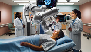
A modern hospital room featuring a robotic arm performing a guided biopsy on a middle-aged Hispanic female patient. Three Hispanic doctors closely observe the procedure, highlighting the advanced technology and sampling precision in oncological pathology.

Robotic Arm-Guided Biopsies: Enhancing Sampling Precision in Oncological Pathology

Robot-assisted biopsies enhance accuracy and reduce complications in oncological pathology.

Read more

Modern medical imaging room featuring a large screen displaying an image fusion of CT and MRI scans, highlighting anatomical structures in vibrant colors. Three physicians, two women and one man, are engaged in a focused discussion in front of the screen. The setting reflects a state-of-the-art hospital in Spain, emphasizing diagnostic accuracy and innovative procedure planning in medical imaging.

Image Fusion of CT and MRI: Enhancing Diagnostic Accuracy and Procedure Planning for Medical Professionals

The fusion of CT and MRI images enhances diagnostic accuracy and optimizes the planning of medical procedures.

Read more
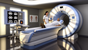
Modern hospital room featuring a PET-CT scanner at the center. A middle-aged Hispanic patient lies on the scanner bed, prepared for the procedure. A Hispanic technician explains the process with a smile. The well-lit room conveys a calm and professional atmosphere. In the background, medical charts and a monitor displaying a 3D image of a tumor highlight advanced technology in oncology for the early detection of tumors.

PET-CT Technology in Oncology: Nuclear Medicine for Early Tumor Detection

PET-CT technology is revolutionizing oncology by enhancing early detection and treatment planning for tumors.

Read more
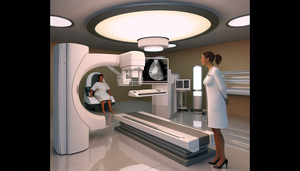
Modern medical imaging room featuring a 3D mammography machine as the focal point. A Hispanic radiologist attentively examines a 3D mammography image on the screen. In the background, a middle-aged Hispanic patient sits comfortably. This scene highlights advanced imaging technology and a professional environment for breast cancer detection and early diagnosis.

3D Mammography: Innovative Imaging Technology for Early Breast Cancer Detection

3D mammography enhances early detection of breast cancer while reducing false positives and anxiety.

Read more
Modern medical imaging room featuring a state-of-the-art PET-CT scanner. A Hispanic physician in his 40s, wearing a white coat and glasses, carefully examines a digital screen displaying PET-CT results, highlighting metabolic activity in the brain and torso. A Hispanic female patient in her 30s, dressed in a hospital gown, is seated on the bed, engaging in conversation with the physician. This scene emphasizes advanced nuclear medicine technology and human interaction in functional studies and scintigraphy diagnostics.

Scintigraphy and Nuclear Medicine: The Role of PET-CT in Functional Studies

Discover how PET-CT is revolutionizing nuclear diagnostics by combining structural and functional data.

Read more

Hispanic scientist in a modern laboratory, examining a clear liquid test tube, symbolizing liquid biopsy. Advanced laboratory equipment and screens displaying graphs and DNA sequences in the background, representing tumor markers. An innovative environment for early cancer detection and diagnosis.

Liquid Biopsy and Tumor Markers: Pioneering Early Detection in Cancer Diagnosis

Liquid biopsy revolutionizes oncological diagnosis by enabling early detection and non-invasive monitoring.

Read more

A modern medical laboratory featuring a diverse team of doctors and scientists collaborating around a digital screen displaying colorful graphs and molecular structures. A Hispanic doctor in his 40s, wearing a white coat and glasses, points to the screen with a pen, explaining the data to his colleagues. This scene highlights innovation and teamwork in the context of early diagnosis, biomarkers, and predictive medicine for effective disease screening.

Biomarkers and Predictive Medicine: Achieving Early Diagnosis and Effective Disease Screening

Discover how biomarkers are revolutionizing early diagnosis and screening of diseases in predictive medicine.

Read more

A middle-aged Hispanic oncology patient smiles while sitting in a comfortable chair in a naturally lit hospital room. She is surrounded by a diverse team of healthcare professionals, including a Caucasian physician, an African American nurse, and an Asian physical therapist, all engaged in a warm discussion. This scene emphasizes the significance of a multidisciplinary approach in comprehensive oncology patient management and palliative care.

Oncology Patient Management and Palliative Care: The Importance of a Multidisciplinary Approach for Comprehensive Care

Multidisciplinary approaches in oncology and palliative care significantly enhance patient quality of life.

Read more

A Hispanic female doctor in her 40s, wearing a white coat, intently examines a digital screen displaying a 3D model of a DNA helix and molecular structures, alongside data analysis graphs and AI algorithms. The scene illustrates a modern precision oncology environment, emphasizing the role of AI-guided therapy and genetic testing in targeted drug treatment response for personalized cancer care.

Targeted Drugs and Treatment Response: How AI-Guided Therapy Enhances Precision Oncology

AI enhances precision oncology by improving treatment responses to targeted therapies.

Read more

Hispanic scientist in a modern laboratory analyzing genetic sequences on a digital screen, surrounded by advanced sequencing machines and diverse colleagues, showcasing collaboration in cutting-edge genomic research with a focus on genetic analysis, massive sequencing, and AI in genomics for the doctor of the future.

AI in Genetic Analysis: From Massive Sequencing to Clinical Interpretation for the Doctor of the Future

AI in genomics is transforming big data into personalized and effective treatments for the future of medicine.

Read more

A modern hospital room equipped with advanced technology, where a diverse group of healthcare professionals analyzes oncology data on a digital screen. The screen displays colorful graphs symbolizing data analysis and AI integration. In the background, a doctor reviews information on a tablet. The collaborative environment emphasizes interoperability, health standards, and data security in oncology data management.

Oncology Data Management with AI: Ensuring Interoperability and Data Security in the Digital Era

Discover how AI is transforming oncology data management by addressing interoperability and security challenges.

Read more

A modern laboratory featuring a diverse team of scientists collaborating on advanced cancer research. In the foreground, a Hispanic female scientist in her 30s examines a screen displaying data visualizations and molecular structures. Beside her, a South Asian male scientist in his 40s points to a 3D model of a DNA strand on a tablet. In the background, a Caucasian female scientist in her 50s analyzes test tubes filled with colorful liquids. The lab is equipped with cutting-edge technology, including monitors showcasing AI algorithms analyzing genetic biomarkers, highlighting an environment of innovation and teamwork in translational medicine and big data analysis.

Identifying Genetic Biomarkers with AI: Accelerating Advanced Cancer Research through Big Data Analysis

AI revolutionizes biomarker identification, enhancing personalized treatments in advanced cancer.

Read more
A Hispanic oncologist in her 40s, wearing a white coat and stethoscope, consults a virtual assistant on her laptop alongside a middle-aged Hispanic patient in a modern, bright hospital room. This interaction emphasizes the role of digital oncology and medical chatbots in enhancing patient support and oncology education.

Virtual Assistants in Oncology: Enhancing Patient Support and Education with Medical Chatbots

Discover how virtual assistants and medical chatbots are transforming support and education in oncology.

Read more

A Hispanic female doctor in her 40s, wearing a white coat and stethoscope, examines a 3D model of the human body on a tablet, symbolizing the use of AI in oncological follow-up. A Hispanic male patient in his 50s, sitting on a hospital bed, watches the doctor with a hopeful smile. The modern, well-lit room reflects an environment of comprehensive care and advanced technology, highlighting the importance of patient monitoring and therapeutic adherence through medical apps.

Oncological Follow-Up with AI: Personalized Patient Monitoring to Enhance Therapeutic Adherence and Comprehensive Care

AI and medical apps personalize oncological follow-up, enhancing adherence and comprehensive care.

Read more

A modern medical laboratory equipped with advanced technology, where a diverse group of physicians, including a Hispanic man and an Asian woman, analyzes complex data on a digital screen related to early cancer detection. The screen displays colorful graphs symbolizing the integration of big data analysis and machine learning in oncological screening. The environment emphasizes innovation and precision in patient evolution and early cancer detection.

Early Cancer Detection: From Oncological Screening to Big Data Analysis and Machine Learning in Patient Evolution

AI in oncology enhances early cancer detection and personalizes treatments for patients.

Read more

A diverse team of physicians analyzes complex data on a digital screen regarding personalized oncological therapies, while a Hispanic patient watches with hope from her bed in a modern hospital room. The image emphasizes collaboration and the use of artificial intelligence in precision oncology, highlighting decision algorithms and targeted treatments for improved response prediction.

Personalized Therapies and AI: Decision Algorithms for Optimal Oncological Treatments in Precision Oncology

Artificial intelligence is transforming precision oncology by optimizing the selection of personalized treatments.

Read more
A modern medical laboratory featuring a diverse team of scientists and physicians, including a Hispanic man and an Asian woman, gathered around a computer screen displaying a 3D model of a tumor with tumor biomarker data. The environment reflects collaboration and innovation, equipped with cutting-edge technology and shelves filled with medical books and journals, emphasizing the importance of tumor classification, molecular analysis, and personalized diagnosis through machine learning.

Tumor Classification with Machine Learning: Advancing Personalized Diagnosis through Molecular Analysis and Biomarkers

AI and tumor biomarkers are revolutionizing oncological diagnosis towards a personalized approach.

Read more

A diverse group of physicians, including a Hispanic oncologist, an Asian radiologist, and a Black data scientist, engage in a discussion in front of a digital screen displaying a 3D model of a tumor along with data and graphs from predictive algorithms. In the background, a Hispanic patient observes from a hospital bed. This scene embodies innovation and optimism in cancer prognosis, highlighting advanced technology and a warm environment, emphasizing the role of predictive algorithms in oncological survival and treatment personalization through cancer big data.

AI in Cancer Prognosis: Predictive Algorithms for Oncological Survival and Treatment Personalization Using Big Data

Artificial intelligence is transforming oncology by personalizing treatments and improving cancer survival rates.

Read more

Modern hospital room featuring an advanced design PET-CT scanner at the center. A diverse team of medical professionals, including a Hispanic physician and an Asian technician, engage in discussion while reviewing detailed PET-CT images on a digital screen. The atmosphere embodies collaboration and innovation in nuclear medicine, highlighting the role of machine learning in advanced diagnosis and functional imaging.

Advancements in Nuclear Medicine: Enhancing PET-CT and Functional Imaging with Machine Learning

AI in nuclear medicine enhances diagnostic accuracy and treatment personalization in PET-CT.

Read more
A Hispanic female radiologist in her 40s, wearing a white coat, carefully examines a digital screen displaying a detailed 3D scan of a human torso, highlighting areas of potential tumors. The modern radiology room is equipped with advanced technology, reflecting a professional approach to early cancer detection through AI diagnosis and precision in oncology.

Oncological Radiology: AI for Early Cancer Detection and Precision in Oncology

Artificial intelligence is revolutionizing oncological radiology, enhancing early detection and diagnostic accuracy.

Read more

A middle-aged Hispanic doctor in a white coat and stethoscope attentively explains a medical chart related to neuroendocrine tumor prognosis to a Hispanic female patient in her 50s in a well-lit hospital consultation room. The desk features medical books, a laptop, and a model of the human endocrine system. The atmosphere is professional and warm, highlighting the supportive relationship between the physician and patient, emphasizing the importance of understanding NET survival and endocrine prognostic factors.

Neuroendocrine Tumors: Prognostic Factors and Life Expectancy in Neuroendocrine Carcinoma Survival

Explore how prognostic factors enhance the survival and treatment of neuroendocrine tumors.

Read more

A middle-aged Hispanic man in a medical consultation, reviewing a medical chart with a Hispanic doctor who is calmly explaining. The scene reflects a supportive doctor-patient relationship, relevant to the prognosis and life expectancy of Merkel cell carcinoma.

Merkel Cell Carcinoma: Prognosis, Survival, and Key Factors in This Rare Skin Cancer

Merkel cell carcinoma is aggressive, but comprehensive treatment significantly improves survival.

Read more

A middle-aged Hispanic doctor in a white coat and stethoscope attentively explains a medical chart to a Hispanic female patient in her 40s in a hospital consultation room. The casually dressed patient listens with interest and concern. Behind them, an anatomical poster details human adipose tissue, highlighting areas where liposarcomas may develop. The scene reflects a professional and compassionate environment, emphasizing the importance of discussing liposarcoma prognosis and survival factors in fat tissue tumors.

Liposarcoma: Prognosis, Survival, and Key Factors in Fat Tissue Tumors

Explore how histological subtypes and the location of liposarcoma impact prognosis and survival.

Read more

A diverse group of healthcare professionals in a modern hospital setting. In the foreground, a Hispanic male physician in his 40s explains a medical chart to a Hispanic female doctor in her 30s. In the background, a smiling Hispanic pediatric patient is seen in her hospital bed, accompanied by a family member. The image conveys an atmosphere of hope and collaboration in the treatment of rhabdomyosarcoma, highlighting the importance of understanding prognostic factors for improved pediatric tumor survival and life expectancy in soft tissue sarcoma.

Rhabdomyosarcoma: Prognosis and Prognostic Factors Influencing Survival in Pediatric Soft Tissue Sarcomas

Explore how genetic factors and emerging treatments influence rhabdomyosarcoma survival.

Read more

Serene hospital consultation room with natural lighting. A middle-aged Hispanic physician with a friendly expression explains a medical chart related to leiomyosarcoma prognosis to a Hispanic female patient in her 40s. The desk is equipped with medical books, a laptop, and a human torso model. In the background, a window reveals a sunny garden, symbolizing hope and tranquility, while emphasizing the importance of understanding soft tissue sarcoma life expectancy and survival rates.

Leiomyosarcoma: Prognostic Factors and Life Expectancy in Soft Tissue Sarcomas

Discover how new therapeutic combinations and genomic models enhance the prognosis of leiomyosarcoma.

Read more

A Hispanic boy of approximately 10 years smiles in a pediatric oncology clinic, accompanied by a Hispanic doctor who speaks to him with empathy. The image reflects a warm and hopeful environment, highlighting compassionate care in the treatment of Ewing sarcoma, emphasizing prognosis, survival, and key prognostic factors for childhood bone tumors.

Ewing Sarcoma: Prognosis, Survival, and Life Expectancy in Childhood Bone Tumors

Explore how multidisciplinary therapy enhances survival in pediatric Ewing sarcoma.

Read more

A middle-aged Hispanic physician in a white coat and stethoscope explains a diagnosis to a Hispanic female patient in her 40s, pointing to a 3D model of a human skeleton on a digital tablet, highlighting the cartilage areas. The patient listens attentively, showing understanding. The setting is professional and empathetic, emphasizing the importance of doctor-patient communication in understanding conditions such as chondrosarcoma, its prognosis, and factors affecting cartilage tumor life expectancy and bone sarcoma survival.

Chondrosarcoma Prognosis: Survival Factors and Life Expectancy in Cartilage Tumors

The management of chondrosarcoma depends on the tumor grade and the presence of metastasis.

Read more

A hopeful 15-year-old Hispanic patient sitting on a hospital bed attentively listens to a Hispanic doctor explaining his osteosarcoma treatment. The room is filled with natural light, creating a warm and optimistic atmosphere, reflecting the importance of understanding osteosarcoma prognosis and bone tumor life expectancy for improved bone sarcoma survival.

Osteosarcoma: Prognosis, Life Expectancy, and Survival Factors in Orthopedic-Oncological Management

Explore how novel biomarkers and targeted therapies can enhance survival in osteosarcoma.

Read more

A middle-aged Hispanic doctor in a white coat and stethoscope carefully examines a brain MRI on a lightbox, focusing on the pituitary gland area. In the background, a Hispanic female patient in her 30s watches the doctor from an examination table. The image conveys professionalism and hope in the context of pituitary cancer prognosis, life expectancy of pituitary tumors, and survival rates for pituitary adenomas.

Pituitary Cancer: Prognosis, Survival Rates, and Key Factors for Pituitary Tumors

Discover how treatment at specialized centers enhances survival in complex pituitary tumors.

Read more

A middle-aged Hispanic physician in a white coat and stethoscope explains a diagnosis to a Hispanic female patient in her 40s, pointing to an anatomical chart of the endocrine system that highlights the adrenal glands. The patient listens attentively in a well-lit medical consultation room, symbolizing a supportive doctor-patient relationship while emphasizing the importance of understanding adrenal cancer prognosis and survival of adrenal tumors.

Adrenal Cancer: Prognosis, Life Expectancy, and Key Prognostic Factors for Adrenal Tumors

Discover how new biomarkers enhance prognosis and survival in adrenal cancer.

Read more

A middle-aged Hispanic physician in a white coat and stethoscope carefully reviews a medical record on a tablet in an office. In the background, an anatomical diagram of the human neck highlights the parathyroid glands. On the desk, there are medical books and a model of the thyroid and parathyroid glands, creating an atmosphere of expertise and care, relevant to parathyroid cancer prognosis and life expectancy in parathyroid carcinoma.

Parathyroid Cancer: Prognosis, Life Expectancy, and Endocrine Survival Factors

Explore how surgery and biomarkers enhance survival in parathyroid cancer.

Read more

A diverse group of healthcare professionals, including a Hispanic female doctor, an Asian male doctor, and a Black nurse, discussing thyroid cancer prognosis and life expectancy based on histological subtypes, with images of thyroid histology on a tablet and anatomical models of the thyroid gland in the background.

Thyroid Cancer Prognosis and Life Expectancy Based on Histological Subtype: Key Factors for Survival

Discover how the histological subtype of thyroid cancer influences prognosis and survival.

Read more

Image of a medical research laboratory featuring three Hispanic doctors discussing in front of a digital screen displaying a 3D model of the human brain, emphasizing the central nervous system. This professional and scientific setting highlights the study of ependymoma prognosis, life expectancy in CNS tumors, and neurological prognostic factors related to ependymoma survival.

Ependymoma: Prognostic Factors and Survival Outcomes in CNS Tumors

Molecular subclassification of ependymoma enhances survival prediction and treatment personalization.

Read more

A Hispanic female doctor in her 40s, wearing a white coat, shows a brain MRI image on a tablet to an 8-year-old Hispanic boy in a hospital bed. The interaction conveys trust and optimism in a warm and welcoming hospital environment, highlighting the importance of understanding medulloblastoma prognosis and childhood brain tumor life expectancy.

Medulloblastoma: Prognosis, Survival, and Life Expectancy in Childhood Brain Tumors

Discover how molecular classification of medulloblastoma enhances prognosis and survival in pediatric patients.

Read more
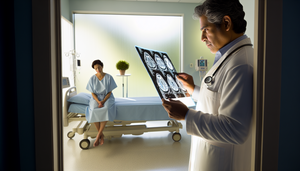
A middle-aged Hispanic physician carefully examines a brain MRI in a serene and well-lit hospital room. The image highlights an area indicating an astrocytoma, a type of glial tumor. In the background, a Hispanic female patient in her 30s sits on the hospital bed, holding a potted plant, symbolizing hope and growth in the context of astrocytoma prognosis and survival.

Astrocytoma: Prognostic Factors and Life Expectancy in Glial Tumors Survival

Discover how genetic mutations and tumor grade impact the survival of astrocytomas.

Read more

A middle-aged Hispanic patient with a serene expression sits on the edge of a hospital bed, gazing at a garden through a window. A Hispanic female doctor, dressed in a white coat and stethoscope, offers support by placing a hand on his shoulder. This scene conveys compassion and hope in a modern hospital setting, highlighting the importance of emotional support in glioblastoma prognosis and brain cancer survival.

Glioblastoma: Prognosis, Life Expectancy, and Survival Factors in Aggressive Brain Cancer

Explore how MGMT promoter methylation and surgical resection enhance survival in glioblastoma.

Read more

A middle-aged Hispanic patient sitting on the edge of a hospital bed gazes out the window with a calm determination. A Hispanic female doctor in a white coat stands beside him, warmly explaining something. The scene illustrates the doctor-patient relationship in the context of multiple myeloma prognosis and life expectancy, highlighting the importance of understanding plasma cell neoplasms survival and bone marrow prognostic factors.

Multiple Myeloma: Prognosis and Life Expectancy Based on Clinical Prognostic Factors

Explore how advancements in treatment and diagnosis are improving prognosis and life expectancy in multiple myeloma.

Read more

A diverse group of healthcare professionals in a modern hospital. In the foreground, a Hispanic female doctor reviews a medical record, while an Asian male doctor points to a digital screen displaying a blood cell diagram, symbolizing acute lymphoblastic leukemia. In the background, a Caucasian nurse interacts with a young patient, creating an atmosphere of care and professionalism. The image conveys hope and collaboration in the treatment of acute leukemia, highlighting the importance of understanding ALL prognostic factors for improving life expectancy and survival rates.

Acute Lymphoblastic Leukemia: Prognosis, Risk Factors, and Survival Rates in Pediatric and Adult Patients

Explore how advancements in personalized therapies are enhancing survival rates in acute lymphoblastic leukemia.

Read more

Middle-aged Hispanic patient in a modern medical consultation, discussing chronic myeloid leukemia (CML) prognosis with a Caucasian doctor. Both are focused on a hopeful prognosis, surrounded by medical charts and a cellular model on the table. The scene reflects a professional and empathetic environment, emphasizing CML life expectancy and survival factors.

Chronic Myeloid Leukemia: Prognosis Evaluation and Life Expectancy Factors for Improved CML Survival

Discover how tyrosine kinase inhibitors have transformed the prognosis of chronic myeloid leukemia, bringing patients' life expectancy closer to normalcy.

Read more

A Hispanic physician in his 40s, wearing a white coat and glasses, examines chromosomal karyotype images in a modern medical laboratory. In the background, there are shelves filled with medical books and a computer screen displaying a 3D model of DNA. The image conveys an advanced research environment focused on acute myeloid leukemia prognosis, highlighting cytogenetic and molecular factors that influence AML life expectancy and acute leukemia survival.

Acute Myeloid Leukemia: Prognosis, Survival, and Key Cytogenetic and Molecular Factors Affecting AML Life Expectancy

The integration of cytogenetic and molecular factors enhances risk stratification and personalizes treatments in AML.

Read more

A middle-aged Hispanic doctor in a white coat and stethoscope calmly explains to a Hispanic patient in a naturally lit hospital room. The patient, sitting on the bed, listens attentively and with hope. The scene conveys an atmosphere of care and optimism, relevant to the prognosis and life expectancy of non-Hodgkin lymphoma, highlighting important hematologic-oncological prognostic factors.

Non-Hodgkin Lymphoma: Prognosis, Life Expectancy, and Survival Rates Across Subtypes

The prognosis and life expectancy in non-Hodgkin lymphoma depend on the specific subtype and prognostic factors.

Read more

A Hispanic male doctor in his 40s, wearing a white coat and stethoscope, calmly explains Hodgkin lymphoma to a Hispanic female patient in her 30s, seated on a hospital bed. The doctor holds a tablet displaying medical charts, while the patient listens attentively. This interaction embodies hope and understanding regarding Hodgkin lymphoma prognosis, life expectancy, and key prognosis factors in hematology-oncology.

Hodgkin Lymphoma: Prognosis, Risk Factors, and Life Expectancy in Hematology-Oncology

Advancements in treatment have significantly improved the prognosis and survival rates of Hodgkin lymphoma.

Read more
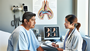
Middle-aged Hispanic patient in a medical examination room discussing with a Hispanic doctor. The doctor, wearing a white coat and stethoscope, takes notes while listening. An anatomical chart of the paranasal sinuses on the wall emphasizes the significance of prognosis and survival in nasal cavity cancer, highlighting key prognostic factors for sinus tumors.

Prognostic Factors and Survival Rates in Nasal Cavity Cancer: Insights on Sinus Tumors and Life Expectancy

Explore how endoscopic techniques and advanced therapies enhance survival in sinus cancer.

Read more

A diverse group of healthcare professionals, including doctors and nurses, gathers around a conference table in a modern hospital. They are discussing nasopharyngeal cancer prognosis while observing a digital screen displaying an anatomical illustration of the nasopharynx and graphs related to nasal tumor survival and prognostic factors. The image reflects a collaborative environment focused on global medical research.

Nasopharyngeal Cancer: Prognostic Factors and Survival Projections for Improved Life Expectancy

Discover how molecular biomarkers and advanced techniques enhance the prognosis of nasopharyngeal cancer.

Read more

A middle-aged Hispanic patient in a modern medical consultation, discussing with a Hispanic otolaryngologist. The doctor, wearing a white coat, holds a clipboard while explaining something to the patient. On the desk, there is a model of the human larynx and medical charts, emphasizing a professional and supportive environment. This scene highlights the importance of understanding laryngeal cancer prognosis, life expectancy, and prognostic factors in head and neck tumors.

Laryngeal Cancer: Prognosis, Life Expectancy, and Survival Factors for Head and Neck Tumors

Explore how early diagnosis and surgical intervention enhance survival rates in laryngeal cancer.

Read more

Middle-aged Hispanic man in a medical consultation, showing concern while listening to a Hispanic female doctor who explains with a reassuring expression. The doctor, wearing a white coat and stethoscope, is in an office with anatomical models of the throat, emphasizing the importance of communication in pharyngeal cancer prognosis, life expectancy, and treatment.

Pharyngeal Cancer: Prognosis, Survival Rates, and Key Prognostic Factors for Oropharyngeal Tumors

Discover how tumor stage and HPV status impact survival in pharyngeal cancer.

Read more

Middle-aged Hispanic patient in a modern medical consultation, discussing oral cancer treatment with a diverse healthcare team. The oncologist presents a 3D model of the oral cavity on a tablet, while the surgeon explains the treatment plan. The nurse is taking notes. The environment is professional and welcoming, emphasizing an optimistic multidisciplinary approach to oral cancer prognosis and survival.

Oral Cancer: Prognosis, Life Expectancy, and Survival Factors in Multidisciplinary Treatment

A multidisciplinary approach and biomarkers such as miR-21 and miR-31 enhance survival in oral cancer.

Read more

Serene medical office with natural lighting. In the foreground, a Hispanic female doctor reviews a medical file, surrounded by medical books and a model of the female reproductive system on her desk, highlighting a focus on gynecological health. In the background, a window reveals a garden, symbolizing hope and life, while emphasizing the importance of understanding vaginal cancer prognosis, life expectancy, and prognostic factors in gynecological neoplasms survival.

Vaginal Cancer: Prognosis, Life Expectancy, and Key Survival Factors for Gynecological Specialists

Discover how prognostic factors like HPV and p16 enhance survival in vaginal cancer.

Read more

Middle-aged Hispanic female doctor in her medical office reviewing data on endometrial cancer prognosis and survival rates on her computer. She is wearing a white coat and a stethoscope, with a poster of the female reproductive system in the background. The scene conveys professionalism and dedication to gynecological oncology, focusing on prognostic factors for endometrial cancer and uterine cancer life expectancy.

Endometrial Cancer: Prognosis, Survival Rates, and Key Prognostic Factors in Clinical Practice

Molecular classification and personalized surgery enhance prognosis and survival in endometrial cancer.

Read more

Hispanic gynecologist reviewing a medical file in her office, with an anatomical diagram of the female reproductive system on the wall and a monitor displaying a graph on cervical cancer prognosis. The scene conveys professionalism and hope, highlighting key aspects of cervical cancer life expectancy and prognostic factors in cervical neoplasia.

Cervical Cancer Prognosis and Life Expectancy: Key Insights for Gynecologists on Survival and Prognostic Factors

Discover how advancements in predictive models and treatments are enhancing survival rates in cervical cancer.

Read more

A middle-aged Hispanic woman with a colorful headscarf sits by a hospital window, gazing outside with a hopeful expression. Soft sunlight gently illuminates her face. Behind her, a reassuring Hispanic doctor smiles while holding a medical record. This scene conveys warmth, support, and optimism, reflecting the importance of understanding ovarian cancer prognosis, life expectancy, and survival factors in gynecological cancer.

Ovarian Cancer: Prognosis, Life Expectancy, and Key Survival Factors in Gynecological Cancer

Discover how stage, histological type, and biomarkers influence ovarian cancer survival.

Read more

A middle-aged Hispanic patient discusses with a Hispanic doctor in a modern medical consultation. The doctor, wearing a white coat and stethoscope, uses a digital tablet to explain important information. The patient listens attentively, showing concern but also hope. This image reflects a supportive and understanding doctor-patient relationship, relevant to the prognosis and life expectancy of penile cancer, as well as the survival of genital tumors and their prognostic factors.

Penile Cancer: Prognostic Factors and Life Expectancy in Clinical Management

Early detection and centralized treatment significantly improve survival in penile cancer.

Read more

A young Hispanic patient, dressed casually, sits with an optimistic expression in a modern medical office. In front of him, a middle-aged Hispanic doctor in a white coat and glasses explains something with a reassuring smile while holding a tablet displaying a graph. The office is bathed in natural light, creating a warm and professional atmosphere, reflecting the hopeful outlook on testicular cancer prognosis and survival rates.

Testicular Cancer: Prognosis, Survival Rates, and Key Factors Influencing Life Expectancy

Testicular cancer has a favorable prognosis with high survival rates, even in advanced stages.

Read more

A middle-aged Hispanic patient with a thoughtful expression sits on a hospital bed, gazing out of a large window that lets in natural light. Beside him, a Hispanic doctor in a white coat holds a clipboard and smiles kindly, conveying support and reassurance. The scene reflects an atmosphere of calm and hope in the context of renal cell carcinoma prognosis, kidney cancer life expectancy, and survival factors.

Renal Cell Carcinoma: Prognosis, Life Expectancy, and Survival Factors in Kidney Cancer

Discover how advancements in predictive models and technologies are enhancing survival rates in renal cancer.

Read more

A middle-aged Hispanic physician in a white coat and stethoscope attentively explains a chart to a Hispanic female patient in her 50s in a hospital consultation room illuminated by natural light. On the desk, there are medical brochures and a model of the human bladder, while an anatomical poster of the urinary system is visible on the wall. The scene conveys trust and understanding in the context of a discussion about bladder cancer prognosis, life expectancy, and prognostic factors for bladder cancer.

Bladder Cancer Prognosis and Survival: Staging and Risk Factors Impacting Life Expectancy

Discover how bladder cancer staging and risk factors impact survival rates.

Read more

Middle-aged Hispanic patient in a medical consultation discussing prostate cancer prognosis with a Hispanic doctor. The doctor is showing a diagram of the prostate gland on a digital tablet. A poster on the wall illustrates the stages of prostate cancer. The scene reflects a serious discussion about prostate cancer life expectancy and survival at different stages, highlighting important prognostic factors for malignant prostatitis.

Prostate Cancer: Prognostic Factors and Life Expectancy Across Stages

Explore how prognostic factors influence survival in prostate cancer across different stages.

Read more

A middle-aged Hispanic doctor carefully reviews the medical history of a Hispanic female patient in her 50s in a well-lit hospital consultation room. The computer monitor displays a graph related to anal cancer prognosis, survival rates, and prognostic factors for anal tumors, although the details are blurred. This interaction reflects a professional and empathetic environment, highlighting the importance of doctor-patient communication in clinical decision-making regarding anal cancer life expectancy.

Prognosis and Survival in Anal Cancer: Key Prognostic Factors for Clinical Management

Explore how tumor differentiation and nodal status impact survival in anal cancer.

Read more
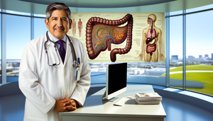
Middle-aged Hispanic doctor in a white coat and stethoscope, carefully examining an anatomical chart of the digestive system with a focus on the gallbladder, in a modern hospital office, highlighting gallbladder cancer prognosis and bile duct cancer survival.

Gallbladder Cancer: Prognosis, Life Expectancy, and Key Survival Factors for Specialists

Explore how advancements in minimally invasive techniques are improving the prognosis of gallbladder cancer.

Read more

A middle-aged Hispanic doctor in a white coat and stethoscope explains an anatomical diagram of the liver on a tablet to a Hispanic female patient in her 40s. The patient listens attentively, showing understanding. The scene reflects professional and compassionate communication about liver cancer prognosis, hepatocellular carcinoma life expectancy, and liver cancer survival.

Liver Cancer: Key Insights into Prognosis, Survival, and Hepatocellular Carcinoma Life Expectancy

Discover how advancements in technology and genomics are enhancing the prognosis and survival rates of hepatocellular carcinoma.

Read more

A middle-aged Hispanic doctor in a white coat and stethoscope explains pancreatic cancer prognosis to a Hispanic female patient in her 50s, using a 3D model on his laptop. The patient listens attentively in a naturally lit hospital room, with an anatomical diagram of the digestive system on the wall, highlighting key prognostic factors for pancreatic cancer and life expectancy.

Pancreatic Cancer: Prognosis, Survival Rates, and Key Prognostic Factors

The integration of biomarkers and molecular factors enhances prognosis and survival in pancreatic cancer.

Read more

A middle-aged Hispanic man in a modern medical consultation, wearing a light blue hospital gown, attentively listens to a Hispanic female doctor explaining the prognosis of esophageal cancer while pointing to a diagram on a screen. The atmosphere is professional and hopeful, highlighting key aspects of esophageal cancer prognosis, life expectancy, and survival factors.

Esophageal Cancer Prognosis and Life Expectancy: Key Prognostic Factors Influencing Survival

Explore how early detection and personalized treatment enhance survival in esophageal cancer.

Read more

A diverse group of medical professionals gathers around a conference table in a modern hospital, seriously discussing gastric cancer prognosis. A middle-aged Hispanic physician points to a chart illustrating the stages of stomach cancer, while others examine an anatomical model of the stomach. The atmosphere is collaborative, focused on improving gastric cancer survival and understanding prognostic factors for stomach cancer.

Gastric Cancer Prognosis and Survival: Evaluating Clinical Stages and Key Prognostic Factors

The integration of biomarkers and novel therapies enhances the prognosis and survival of gastric cancer.

Read more

A diverse group of healthcare professionals in a modern hospital, including a Hispanic physician, a Black nurse, and an Asian medical researcher, discuss in front of an interactive digital screen displaying a 3D model of the human colon. The screen highlights areas related to colorectal cancer, with labels such as "prognostic factors" and "life expectancy." The scene reflects a professional and collaborative environment focused on advancing medical knowledge and patient care, emphasizing colorectal cancer prognosis and survival outcomes.

Colorectal Cancer Prognosis: Key Prognostic Factors and Life Expectancy for Medical Practice

Explore how prognostic factors influence survival and life expectancy in colorectal cancer.

Read more

A middle-aged Hispanic man in a light blue hospital gown attentively listens to a Hispanic female doctor in her modern, clean office. The doctor, with a friendly expression, holds a clipboard while explaining important information about mesothelioma prognosis and life expectancy. The professional and compassionate environment is enhanced by a diagram of the human respiratory system on the wall, highlighting the significance of understanding pleural tumor survival and mesothelioma prognostic factors.

Mesothelioma Prognosis and Life Expectancy: Key Factors for Pleural Tumor Survival

Pleural mesothelioma has an unfavorable prognosis, but early detection and combined therapies can improve survival.

Read more

Image of a middle-aged Hispanic physician in a hospital office, intently reviewing a chart on small cell lung cancer prognosis, survival rates, and prognostic factors. Dressed in a white coat with a stethoscope, a lung scan is displayed on the computer screen, and a lung model is on the desk, reflecting expertise and dedication to the study of SCLC life expectancy and lung cancer survival.

Small Cell Lung Cancer: Survival Evaluation and Prognostic Factors for Improved Life Expectancy

Explore how molecular classification and immunotherapy promise to enhance the prognosis of SCLC.

Read more

Brightly lit hospital room featuring a middle-aged Hispanic patient in bed, looking hopeful. Beside him, a smiling Hispanic doctor holds a tablet displaying a lung scan image, symbolizing an optimistic and supportive approach to non-small cell lung cancer prognosis, NSCLC life expectancy, and lung cancer survival.

Prognosis and Survival in Non-Small Cell Lung Cancer: Current Perspectives on NSCLC Life Expectancy and Prognostic Factors

Explore how targeted therapies and immunotherapies are transforming the prognosis of NSCLC.

Read more

Middle-aged Hispanic doctor in a modern medical office, explaining melanoma stages and survival rates to a Hispanic female patient in her 40s, with a chart on his laptop and a brochure titled "Melanoma: Prognosis and Life Expectancy." The scene conveys a professional and hopeful atmosphere, highlighting melanoma prognostic factors and skin cancer survival.

Melanoma Prognosis and Life Expectancy: Understanding Risk Factors and Stages for Improved Skin Cancer Survival

Discover how risk factors and stages of melanoma impact survival and treatment outcomes.

Read more

A middle-aged Hispanic man in a medical consultation, attentively observing a Hispanic female doctor holding a digital tablet displaying medical statistics. The scene reflects a professional and collaborative environment, relevant to squamous cell carcinoma prognosis, life expectancy, and survival rates.

Prognosis and Life Expectancy in Squamous Cell Carcinoma: Key Factors for Effective Skin Cancer Survival

Early detection and appropriate treatment are crucial for improving survival in squamous cell carcinoma.

Read more

A scene in a modern medical office featuring a middle-aged Hispanic female doctor, smiling and exuding optimism as she reviews an upward trend graph on her laptop. In front of her, a Hispanic female patient in her 50s listens attentively. Natural light fills the room, which is adorned with plants and a pink ribbon poster, a symbol of breast cancer awareness. The image conveys professionalism, empathy, and hope in the context of breast cancer prognosis, survival, and prognostic factors.

Breast Cancer Prognosis and Life Expectancy: Key Prognostic Factors for Clinical Practice

Explore how key factors and prognostic tools personalize breast cancer treatment.

Read more

A Hispanic physician in his 40s, wearing a white coat, carefully examines a digital tablet displaying a DNA sequence in a modern laboratory focused on personalized medicine and pharmacogenomics. In the background, a large screen showcases a 3D model of a DNA double helix, symbolizing genetic research. The environment reflects advanced technology and innovation in precision medicine, highlighting the importance of biomarkers and targeted therapies.

Personalized Medicine and Pharmacogenomics: Advancing Targeted Therapies through Biomarkers and Precision Medicine

Discover how personalized medicine and pharmacogenomics are transforming medical treatments into more effective and safer therapies.

Read more

A diverse group of healthcare professionals discusses cancer screening in a medical office. In the foreground, a Hispanic female doctor in her 40s points to cancer screening statistics on a digital screen, while an African American male doctor in his 50s listens attentively. In the background, a Caucasian female nurse in her 30s works on a computer next to brochures titled "Cancer Screening." The scene reflects a collaborative effort in the early detection of precancerous lesions through effective diagnostic tests.

Effective Cancer Screening Practices: Early Detection of Precancerous Lesions and Diagnostic Tests

Discover how advanced cancer screening can reduce mortality through early detection of cancer.

Read more

A diverse group of physicians discusses in front of a digital screen displaying 3D models of cancer cells and DNA strands, symbolizing advances in oncology, immunotherapy, and precision medicine in a modern, technology-driven hospital environment.

Advances in Oncology: The Role of Immunotherapy and Precision Medicine in Oncological Treatments and Biomarkers

Discover how immunotherapy and precision medicine are revolutionizing personalized oncology treatments.

Read more

A diverse team of radiologists examines a high-resolution 3D image of a human organ on a digital screen, featuring overlays of data and analysis generated by AI, symbolizing the integration of artificial intelligence in emerging diagnostic technologies. In the background, a technician prepares a liquid biopsy sample in a modern, technologically advanced hospital setting.

Emerging Diagnostic Technologies: Liquid Biopsy and AI in Radiology for Medical Innovation

Liquid biopsy and AI in radiology are revolutionizing oncological diagnosis with enhanced precision and personalization.

Read more

A middle-aged Hispanic woman attentively examines a computer screen displaying a detailed magnetic resonance imaging scan, highlighting an area of interest in a muscle mass potentially indicative of leiomyosarcoma. The modern medical office is well-lit, featuring contemporary equipment and a welcoming atmosphere.

Leiomyosarcoma: Identifying Muscle Masses and the Importance of MRI for Early Detection

Magnetic resonance imaging is crucial for the early detection and effective treatment of leiomyosarcoma.

Read more

A Hispanic 10-year-old boy sitting on a hospital stretcher, displaying discomfort and holding his leg, while a Hispanic doctor examines his leg, illustrating the importance of recognizing fever and bone pain in the early diagnosis of Ewing sarcoma in children, along with elevated LDH and imaging studies.

Ewing Sarcoma: Recognizing Fever and Bone Pain in Children for Rapid Diagnosis

Early detection of Ewing sarcoma improves prognosis by recognizing persistent fever and bone pain.

Read more
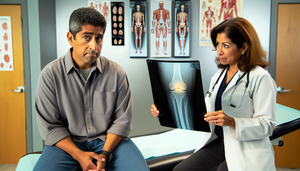
A middle-aged Hispanic man, visibly concerned, is seated in a medical office. In front of him, a Hispanic female doctor in her 40s holds a magnetic resonance imaging scan that highlights an area in the cartilage, suggesting a possible chondrosarcoma. This scene emphasizes the importance of early orthopedic diagnosis and guided biopsy in addressing persistent pain.

Chondrosarcoma: Persistent Pain and the Crucial Role of Guided Biopsy in Early Orthopedic Diagnosis

Guided biopsy is crucial for the early diagnosis of chondrosarcoma and improving prognosis.

Read more
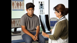
A 15-year-old Hispanic male in a modern medical examination room, with a Hispanic female doctor showing a bone X-ray that suggests possible osteosarcoma. The doctor explains the results, emphasizing the significance of early detection, bone pain assessment, and the role of a bone biopsy in diagnosis.

Early Detection of Osteosarcoma in Young Patients: Importance of Bone X-ray and Biopsy for Bone Pain and Local Inflammation

Early detection of osteosarcoma in young patients improves prognosis through radiographic evaluation and biopsy.

Read more

A Hispanic physician in his 40s, wearing a white coat and glasses, carefully examines magnetic resonance imaging scans of the sellar region, highlighting the pituitary gland for signs of pituitary cancer. In the background, a Hispanic female patient in her 30s observes calmly. This image emphasizes the importance of early detection and advanced imaging techniques in diagnosing visual disturbances and other early symptoms related to hyperprolactinemia.

Pituitary Cancer: Early Symptoms, Visual Disturbances, and Sellar MRI Detection

Early detection of pituitary cancer through sellar MRI enhances clinical prognosis.

Read more
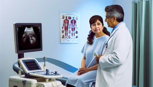
Medical consultation in a modern clinic: a middle-aged Hispanic woman in a blue gown attentively listens to a Hispanic physician pointing at an ultrasound image of a thyroid nodule on a computer screen. The physician holds an ultrasound probe, emphasizing the importance of early diagnosis of thyroid cancer, particularly in cases presenting with dysphagia, and the role of fine needle aspiration (FNA) in evaluation.

Thyroid Cancer: Early Diagnosis of Thyroid Nodules through Cervical Ultrasound and Fine Needle Aspiration (FNA)

Early evaluation of thyroid nodules with ultrasound and fine-needle aspiration enhances the diagnosis of thyroid cancer.

Read more

A Hispanic physician in his 40s, wearing a white coat and stethoscope, carefully examines a brain MRI with contrast on a computer screen, highlighting areas that suggest a possible ependymoma. The desk is cluttered with medical books and handwritten notes, set in a professional and well-lit environment, indicative of a thorough investigation into signs of intracranial hypertension and the need for a tumor biopsy.

Ependymoma: Recognizing Signs of Intracranial Hypertension and Effective Diagnostic Techniques Including MRI with Contrast and Tumor Biopsy

Discover how contrast-enhanced MRI and tumor biopsy are crucial in the diagnosis of ependymoma and its complications.

Read more

A Hispanic boy approximately 6 years old sits on a hospital bed in a modern medical imaging room. He wears a light blue hospital gown and curiously observes an advanced MRI machine. A Hispanic radiologist explains the procedure to him, fostering a supportive and calming environment, emphasizing the importance of early detection of medulloblastoma in pediatric diagnosis, particularly in cases presenting with ataxia and morning vomiting.

Early Detection of Medulloblastoma in Children: The Role of Brain Imaging in Diagnosing Ataxia and Morning Vomiting

Early detection of medulloblastoma in children enhances clinical outcomes through advanced brain imaging techniques.

Read more

An 8-year-old Hispanic boy, slightly pale, sitting on an examination table in a pediatrician's office. A middle-aged Hispanic pediatrician is explaining something to the worried parents of the child. The professional and welcoming environment features posters emphasizing the importance of early detection of pediatric warning signs, including symptoms of acute lymphoblastic leukemia, recurrent infections, lymphadenopathy, and the need for a bone marrow biopsy.

Acute Lymphoblastic Leukemia: Pediatric Warning Signs, Recurrent Infections, and the Importance of Bone Marrow Biopsy

Early detection of acute lymphoblastic leukemia in children improves clinical outcomes through warning signs.

Read more

A Hispanic physician in his 40s, wearing a white coat and glasses, examines chromosomal images on a computer screen in a modern medical laboratory. The images display a karyotype with chromosomal abnormalities associated with chronic myeloid leukemia. The doctor takes notes on a clipboard, emphasizing the importance of cytogenetic analysis in the early diagnosis of this condition, which can lead to chronic fatigue and splenomegaly, and monitor white blood cell count.

Chronic Myeloid Leukemia: Early Identification and the Role of Cytogenetics in Diagnosis, Chronic Fatigue, and Splenomegaly

Early identification of chronic myeloid leukemia through cytogenetics improves prognosis and treatment.

Read more

Middle-aged Hispanic patient in a medical examination room, appearing fatigued and pale, while a Hispanic doctor explains with a medical chart in hand. In the background, a diagram of bone illustrates the significance of doctor-patient communication in diagnosing acute myeloid leukemia, highlighting symptoms such as fatigue, pallor, and the presence of blasts in peripheral blood and bone marrow aspirate.

Acute Myeloid Leukemia: Identifying Non-Specific Symptoms and Confirming Diagnosis with Bone Marrow Aspirate

Discover how to recognize nonspecific symptoms of acute myeloid leukemia and confirm with bone marrow aspiration.

Read more
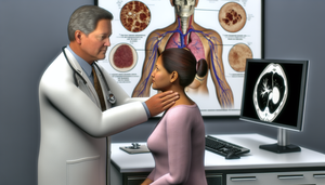
A Hispanic male physician in his 40s, wearing a white coat and stethoscope, carefully examines the lymph nodes in the neck of a Hispanic female patient in her 30s during a medical consultation. In the background, a medical poster illustrates the human lymphatic system. Nearby, a computer screen displays a PET scan image. This scene highlights a thorough medical examination and the use of advanced technology for diagnosing non-Hodgkin lymphoma, particularly in cases presenting with lymphadenopathy and fever of unknown origin.

Early Identification of Non-Hodgkin Lymphoma: Lymphadenopathy, Fever of Unknown Origin, and the Role of PET Scans in Diagnosis

Early identification of lymphadenopathy and the use of PET scans enhance the diagnosis of non-Hodgkin lymphoma.

Read more

A middle-aged Hispanic patient in a light blue hospital gown attentively listens to a Hispanic doctor holding a medical history. On the desk, a model of the lymphatic system highlights the lymph nodes, emphasizing the importance of communication in diagnosing Hodgkin lymphoma, particularly in cases presenting with painless lymphadenopathy and B symptoms like night sweats.

Hodgkin Lymphoma: Painless Lymphadenopathy and the Role of Lymph Node Biopsy in Diagnosis

Explore how lymph node biopsy is crucial for diagnosing Hodgkin lymphoma in the presence of painless lymphadenopathy.

Read more
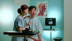
A middle-aged Asian man with a calm yet concerned expression is seated in a well-lit medical examination room, wearing a light blue hospital gown. An Asian female doctor in her 40s, holding a nasofibroscope, is reassuringly explaining the procedure to him. In the background, a 3D model of the nasopharynx is displayed on a computer screen. This image emphasizes the importance of early detection of nasopharyngeal cancer through nasofibroscopy, particularly in patients presenting with nasal obstruction and epistaxis.

Nasopharyngeal Cancer: Recognizing Early Signs, Nasal Obstruction, and the Importance of Timely Nasofibroscopy

Early detection of nasopharyngeal cancer through nasofibroscopy significantly improves prognosis and treatment outcomes.

Read more

A middle-aged Hispanic man in a medical examination room, displaying a slight look of concern, listens to a Hispanic female doctor who is holding a laryngoscope and explaining a procedure. Nearby, a diagram of the larynx is visible on a screen. This interaction emphasizes the critical role of communication in the early diagnosis of laryngeal cancer, particularly in cases presenting with persistent hoarseness and referred otalgia.

Laryngeal Cancer: Recognizing Persistent Hoarseness and Confirming Diagnosis with Direct Laryngoscopy for Early Detection

Persistent hoarseness may be an early sign of laryngeal cancer, diagnosable through direct laryngoscopy.

Read more

A middle-aged Hispanic patient in a dental examination chair, with a Hispanic dentist performing a stomatological examination of the mouth. The scene emphasizes the importance of early detection of oral cancer and the differential diagnosis of oral lesions, including leukoplakia, in a professional and modern setting.

Oral Cancer: Stomatological Examination of Oral Lesions and the Importance of Early Detection in Differential Diagnosis

Early detection of oral cancer enhances survival through innovative diagnostic technologies and biomarkers.

Read more

A Hispanic female doctor in a modern medical office explains a medical procedure to a middle-aged Hispanic patient. The doctor points to the endometrium on a diagram of the female reproductive system, emphasizing the importance of early detection of endometrial cancer, patient education on abnormal uterine bleeding, and endometrial biopsy techniques.

Endometrial Cancer: Early Warning Signs of Abnormal Uterine Bleeding and Biopsy Techniques

Early detection of endometrial cancer improves prognosis through SUA evaluation and biopsies.

Read more

A 25-year-old Hispanic man thoughtfully examines himself in the mirror of a modern bathroom, highlighting the importance of self-examination for the early detection of testicular cancer and the identification of testicular masses, complemented by scrotal ultrasound and tumor markers (AFP, β-hCG).

Testicular Cancer: Self-Examination and Tumor Markers (AFP, β-hCG) for Early Detection of Testicular Mass

Early detection of testicular cancer improves prognosis through self-examination, ultrasound, and tumor markers.

Read more

Forty-year-old Hispanic doctor in a modern hospital examining renal ultrasound and computed tomography images on a large screen. The images reveal a kidney with an abdominal mass indicative of renal cell carcinoma. The doctor points to the mass with a pen, while a Hispanic nurse takes notes on a digital tablet in the background.

Renal Cancer: Ultrasound and CT Findings for Timely Diagnosis of Renal Cell Carcinoma

Timely diagnosis of renal cancer is enhanced by ultrasound and tomography, distinguishing between benign and malignant lesions.

Read more
A middle-aged Hispanic physician in a white coat and stethoscope attentively explains a medical chart to a Hispanic female patient in her 50s in a modern examination room. The patient is seated on an exam table, showing interest. The image emphasizes the importance of early detection of bladder cancer and the diagnosis of initial hematuria, highlighting the role of cystoscopy and urinary cytology in a professional and warm environment.

Bladder Cancer: Early Detection of Initial Hematuria and Diagnostic Techniques

Early detection of hematuria and urinary biomarkers enhances the diagnosis of bladder cancer.

Read more

A middle-aged Hispanic physician in a white coat and stethoscope explains to a Hispanic patient in his 50s the use of a prostate gland model to illustrate the importance of prostate-specific antigen (PSA) levels and digital rectal exams in the early diagnosis of prostate cancer. On the desk, there are charts showing PSA levels, emphasizing the focus on early detection.

Prostate Cancer: The Role of PSA Testing and Digital Rectal Exam in Early Diagnosis

Early detection of prostate cancer improves with the combination of PSA, digital rectal examination, and new technologies.

Read more

A middle-aged Hispanic patient sitting on an examination table attentively listens to a Hispanic doctor holding an anoscope and pointing to a diagram of the digestive system. This scene emphasizes the importance of anoscopy in the early detection of anal cancer, particularly in patients at risk for HPV-related conditions, and highlights symptoms such as rectal bleeding and anal pain.

Early Signs of Anal Cancer: Recognizing Symptoms and the Role of Anoscopy in Diagnosis

Early detection of anal cancer through anoscopy improves clinical outcomes in high-risk patients.

Read more

Medical consultation in a modern clinic where a middle-aged Hispanic female doctor, wearing a white coat and stethoscope, explains a gallbladder diagram on a tablet to a middle-aged Hispanic patient. The patient, seated on an examination table, displays interest and concern. This image illustrates a professional and educational environment, relevant to gallbladder cancer, right upper quadrant pain, cholelithiasis, cholangiography, and the importance of early diagnosis.

Gallbladder Cancer: Recognizing Right Upper Quadrant Pain and Early Diagnosis Methods for Cholelithiasis

Explore how advancements in imaging and biomarkers enhance the early diagnosis of gallbladder cancer.

Read more

A middle-aged Hispanic man in a medical office attentively observes a 3D model of a liver on a computer screen. The doctor, a Hispanic woman in her 40s, points to the model while explaining the importance of early detection of liver cancer, particularly in patients at risk of cirrhosis. The atmosphere is professional and warm, emphasizing patient education on focal liver lesions and the role of alpha-fetoprotein in diagnosis.

Early Identification of Liver Cancer in Patients at Risk of Cirrhosis: The Role of Alpha-Fetoprotein and Liver Ultrasound in Detecting Focal Liver Lesions

Early detection of liver cancer in cirrhotic patients improves prognosis through ultrasound and alpha-fetoprotein.

Read more

A Hispanic physician in his 40s, wearing a white coat and stethoscope, carefully examines a medical file in a modern office. On the desk, there are medical books and a computer displaying a 3D model of the pancreas, highlighting areas affected by pancreatic cancer. In the background, a diagram of the human digestive system with the pancreas labeled is visible. The scene reflects a medical research environment focused on pancreatic cancer, nonspecific abdominal pain, jaundice, CA 19-9, and endoscopic ultrasound.

Pancreatic Cancer: Initial Findings, Tumor Markers, and the Role of Endoscopic Ultrasound in Diagnosis

Discover how the combination of tumor markers and new technologies enhances the early diagnosis of pancreatic cancer.

Read more

Middle-aged Hispanic man sitting in a kitchen, showing concern while touching his throat, suggesting possible progressive dysphagia. On the table, there is a glass of water and an untouched plate of bread, indicating difficulty swallowing, which may be a sign of esophageal cancer. Early detection through esophageal endoscopy and esophagram is crucial.

Esophageal Cancer: Recognizing Progressive Dysphagia for Early Detection and Timely Action

Early detection of progressive dysphagia improves the prognosis of esophageal cancer.

Read more

A Hispanic physician in a white coat explains the digestive system to a middle-aged Hispanic couple using a diagram on a tablet, pointing to the colon area. The modern and well-lit medical consultation room features posters on colorectal cancer awareness, emphasizing the importance of colonoscopy, fecal occult blood tests, and recognizing symptoms like rectal bleeding and polyps.

Colorectal Cancer: Early Detection through Symptom Monitoring and Colonoscopy for Rectal Bleeding and Polyps

Early detection of colorectal cancer through colonoscopy and symptom monitoring reduces mortality.

Read more

A middle-aged Hispanic man in a modern medical consultation, attentively observing a Hispanic female doctor holding a chest X-ray. The doctor points to a specific area in the image, emphasizing the importance of early detection and key tests such as spirometry and lung biopsy in diagnosing small cell lung cancer and its associated respiratory symptoms.

Small Cell Lung Cancer: Early Detection, Key Tests, and Respiratory Symptoms

Discover how advancements in biomarkers and non-invasive techniques are enhancing the detection of small cell lung cancer.

Read more
A Caucasian dermatologist in his 40s examines the arm of a Hispanic female patient in her 30s using a dermatoscope. The patient presents several atypical moles of varying sizes and colors. This image emphasizes the critical role of early diagnosis of skin cancer, particularly melanoma, and the application of the ABCDE criteria for melanoma assessment.

Melanoma: Recognizing Atypical Moles for Early Skin Cancer Diagnosis Using the ABCDE Method

Discover how to identify suspicious moles and enhance early melanoma diagnosis.

Read more

A middle-aged Hispanic man in a dermatology clinic, displaying a concerned yet hopeful expression, as a Hispanic dermatologist examines a light brown skin lesion on his forearm with a dermatoscope. The lesion has irregular borders, suggesting early-stage squamous cell carcinoma. This image emphasizes the importance of clinical evaluation for precancerous lesions like actinic keratosis and the critical role of early detection in preventing metastasis.

Early Detection and Management of Squamous Cell Carcinoma: Clinical Evaluation of Precancerous Lesions and Actinic Keratosis

Early detection and appropriate management of squamous cell carcinoma prevent its invasive progression.

Read more

Hispanic female doctor in a medical office explaining mammography to a Hispanic patient, emphasizing the importance of early breast cancer diagnosis and the initial symptoms. Educational posters about breast cancer are visible in the background, highlighting clinical breast examination and breast ultrasound.

Early Breast Cancer Diagnosis: Key Initial Symptoms and Importance of Clinical Breast Examination, Mammography, and Ultrasound

Early detection of breast cancer improves survival through mammograms, ultrasounds, and new biomarkers.

Read more

Bright medical office where a middle-aged Hispanic doctor explains a medical chart to a young Hispanic patient. The desk is cluttered with medical books and a computer displaying a brain MRI image. The walls are adorned with anatomical posters of the brain and nervous system. This scene emphasizes the importance of early diagnosis and differentiation in conditions such as Von Hippel-Lindau disease, cerebral hemangioblastomas, pheochromocytoma, and the role of genetic screening in neurofibromatosis.

Von Hippel-Lindau Disease: Early Diagnosis, Genetic Screening, and Differentiation from Neurofibromatosis

Early identification and differentiation of VHL and NF1 is crucial for appropriate management.

Read more

Hispanic pediatrician examining a child during a medical consultation, with the child's mother beside them. The scene reflects a professional and warm environment, emphasizing the importance of early diagnosis of Fanconi anemia, congenital malformations, and the differentiation of other bone marrow aplastic conditions, including the DEB test for chromosomal fragility.

Fanconi Anemia: Early Diagnosis and Differentiation from Other Aplastic Bone Marrow Disorders

Discover how early diagnosis of Fanconi anemia enhances prognosis by differentiating it from other bone marrow aplasias.

Read more

Middle-aged Hispanic patient in a modern medical examination room, discussing with a Hispanic doctor who holds a tablet displaying a diagram of the human body. An anatomical poster of the circulatory system is visible on the wall. This scene emphasizes the significance of a comprehensive evaluation in systemic amyloidosis, including cardiac assessment and abdominal fat biopsy for abnormal proteins, crucial for differentiating multiple myeloma.

Systemic Amyloidosis: Evaluating Multisystem Symptoms and Differentiating from Multiple Myeloma

Discover how to differentiate systemic amyloidosis from multiple myeloma through multisystem evaluation and advanced techniques.

Read more

Middle-aged Hispanic doctor in his medical office, reviewing hematological data on a computer. Dressed in a white coat and glasses, symbolizing careful attention in managing azathioprine therapy and monitoring for myelosuppression and hepatic toxicity.

Azathioprine: Adverse Reactions and Hematological Monitoring in Long-Term Immunosuppressant Therapy

Discover how genotyping and hematological monitoring enhance the safety of azathioprine therapy.

Read more

Medical consultation with a middle-aged Hispanic doctor explaining the use of methotrexate, an antirheumatic drug, to a Hispanic patient. The doctor holds a bottle of methotrexate, and there is a brochure about folic acid on the desk. The setting is professional and conveys trust, highlighting the importance of monitoring for hepatotoxicity and mucositis.

Methotrexate: Managing Side Effects, Hepatotoxicity, Mucositis, and the Role of Folic Acid in Antirheumatic Therapy

Discover how folates can mitigate methotrexate toxicity and enhance its tolerance.

Read more

A Hispanic physician in his 40s, wearing a white coat and stethoscope, carefully examines the swollen lymph nodes in the neck of a Hispanic female patient in her 30s in a medical consultation room. The patient, seated on an examination table, displays a look of moderate concern. This image highlights the critical importance of accurate diagnosis to differentiate between lymphoma and lymph node tuberculosis, including the role of biopsy and the identification of acid-fast bacilli.

Lymphoma vs. Tuberculosis: Identifying the Cause of Persistent Swollen Lymph Nodes and the Role of Biopsy in Diagnosing Acid-Fast Bacilli

Accurate differentiation between lymphoma and lymph node tuberculosis is crucial for appropriate patient management.

Read more

A Hispanic male physician in his 40s, wearing a white coat and stethoscope, explains blood test results to a Hispanic female patient in her 30s. The physician points to a chart on a tablet displaying serum iron levels, highlighting "Iron Deficiency Anemia" and "Anemia of Chronic Disease." The patient listens attentively. In the background, there are anatomical posters and a plant, creating a professional and welcoming environment.

Iron Deficiency Anemia vs. Anemia of Chronic Disease: Interpreting Ferritin and Serum Iron Levels in Low MCV Cases

Discover how to differentiate between iron deficiency anemia and anemia of chronic disease by interpreting iron values.

Read more
A middle-aged Hispanic patient on an examination table looks at a monitor displaying endoscopic images of the esophagus: one showing signs of esophageal cancer and the other exhibiting characteristics of achalasia. A Hispanic doctor points to the images, explaining the differences. The setting is professional and educational, with visible medical equipment, highlighting the importance of manometry and endoscopy with biopsy in diagnosing dysphagia.

Esophageal Cancer and Achalasia: Risk Factors and Endoscopic Findings in Dysphagia Evaluation

Explore how endoscopic surveillance in achalasia can prevent esophageal cancer.

Read more

Modern medical consultation with a middle-aged Hispanic doctor discussing lupus treatment options, including corticosteroids, immunosuppressants, and rituximab, with a Hispanic patient. The desk features medical brochures and a model of the immune system, creating a professional and warm atmosphere that reflects trust and care in symptomatic management and organ-specific conditions.

Lupus Treatment: Corticosteroids, Immunosuppressants, and Biological Therapies for Symptomatic Management and Organ-Specific Conditions

Explore how corticosteroids, immunosuppressants, and biological therapies are transforming the treatment of autoimmune diseases.

Read more

A Hispanic physician in his 40s, wearing a white coat and stethoscope, explains a procedure to a Hispanic female patient in her 50s using a model of the human stomach. In the background, an endoscopy machine displays a clear image of the stomach lining on the monitor. The scene reflects a professional and caring environment, emphasizing the importance of doctor-patient communication in the diagnosis of gastric cancer, including adenocarcinoma and the use of tumor markers, as well as the relevance of upper gastrointestinal endoscopy and Borrmann classification.

Gastric Cancer Diagnosis: Upper Gastrointestinal Endoscopy, Biopsy, and Borrmann Classification for Adenocarcinoma Detection

Discover how endoscopy and biopsy are crucial in the diagnosis of gastric cancer.

Read more

A Hispanic physician in his 40s, wearing a white coat and stethoscope, examines abdominal CT images that detail the pancreas. Nearby, a computer screen displays a graph showing rising levels of the CA 19-9 marker. The setting is a modern, well-organized medical office, emphasizing advanced medical analysis and pancreatic cancer diagnosis, including considerations for obstructive jaundice and endoscopic biopsy.

Pancreatic Cancer Diagnosis: The Role of Abdominal CT and CA 19-9 in Obstructive Jaundice Evaluation

The combination of abdominal CT and CA 19-9 is crucial but requires caution to avoid misdiagnoses in pancreatic cancer.

Read more

Medical team in a modern hospital discussing a pancreatic cancer treatment plan. The group includes a surgeon, an oncologist, and a palliative care specialist, all of Hispanic descent, interacting with a patient in a hospital bed. The image highlights the multidisciplinary approach to duodenopancreatectomy and compassionate care, emphasizing the role of gemcitabine in treatment.

Pancreatic Cancer Treatment: Whipple Surgery, Gemcitabine, and a Multidisciplinary Approach to Palliative Care

Explore how Whipple surgery, chemotherapy, and palliative care enhance the treatment of pancreatic cancer.

Read more

Diverse medical team discussing gastric cancer treatment options in a modern hospital. A Hispanic surgeon and an Asian oncologist review a 3D model of the stomach on a laptop. In the background, a Hispanic patient consults with a nutritionist about balanced dietary support. The image emphasizes the importance of multidisciplinary collaboration in gastric cancer treatment, including partial or total gastrectomy and adjuvant chemotherapy.

Gastric Cancer Treatment: Partial or Total Gastrectomy, Adjuvant Chemotherapy, and Nutritional Support

Clinical nutrition is crucial for enhancing recovery and survival following gastrectomy in gastric cancer.

Read more

A Hispanic physician in his 40s, wearing a white coat and stethoscope, reviews computer diagnostic images in a modern medical office. The images display detailed scans of the liver, highlighting areas of interest related to liver cancer diagnosis. On the desk, there are medical books and vials labeled as tumor markers. In the background, medical certificates adorn the wall, and a window offers a view of a sunny day. The scene reflects a professional approach to diagnosing liver cancer, emphasizing the importance of AFP, liver ultrasound, CT, MRI, and underlying cirrhosis.

Liver Cancer Diagnosis: Tumor Markers, Imaging Studies, and the Role of AFP in Cirrhosis

The combination of tumor markers and imaging studies enhances the early detection of liver cancer.

Read more

A diverse team of three physicians discusses colorectal cancer treatment in a modern hospital setting. A Hispanic female doctor holds a tablet displaying a diagram of the digestive system, while an African descent male doctor points to the diagram. An Asian female doctor takes notes on a clipboard. The image reflects a multidisciplinary approach to treatment, highlighting collaboration in surgical resection, the use of oxaliplatin and bevacizumab, and considerations for colostomy.

Colorectal Cancer Treatment: Surgical Resection, Chemotherapy with Oxaliplatin, and Bevacizumab Options

Explore how surgery, chemotherapy, and biological therapies enhance the treatment of colorectal cancer.

Read more

A middle-aged Hispanic physician in a white coat and stethoscope explains the colonoscopy procedure to a Hispanic female patient in her 50s, pointing to an anatomical diagram of the digestive system on a digital screen. The casually dressed patient listens attentively, displaying a slight look of concern. Nearby, medical tools including a colonoscope and labeled vials representing biomarkers are visible. This image emphasizes the significance of colorectal cancer diagnosis, including CEA levels, biopsy, and TNM staging.

Colorectal Cancer Diagnosis: Colonoscopy, CEA Markers, and TNM Staging

Explore how colonoscopy and innovative biomarkers enhance the diagnosis and treatment of colorectal cancer.

Read more

Medical consultation in a modern hospital where a middle-aged Hispanic patient listens to a Hispanic doctor explaining schwannoma treatment options for acoustic neuroma, displaying an anatomical diagram of the ear on a digital tablet. In the background, a monitor shows a brain MRI.

Acoustic Neuroma Treatment: Surgery, Radiotherapy, and Post-Operative Rehabilitation for Schwannoma Management

Explore treatment and rehabilitation options to enhance quality of life after an acoustic neuroma.

Read more

A middle-aged Hispanic doctor in a white coat and stethoscope discusses prostate cancer treatment options, including radical prostatectomy, radiotherapy, and hormone therapy, with an older Hispanic patient. The doctor uses a digital tablet displaying a 3D model of the prostate gland. On the desk, there are medical brochures in Spanish about "Active Surveillance," "Radiotherapy," and "Surgery." The consultation room is modern and well-lit, featuring a window with an urban landscape. This scene illustrates a collaborative relationship between doctor and patient, emphasizing the importance of PSA monitoring in treatment decisions.

Prostate Cancer Treatment: Active Surveillance, Radiotherapy, and Radical Prostatectomy Options

Explore how to tailor prostate cancer treatment with active surveillance, radiation therapy, and surgery.

Read more

A middle-aged Hispanic physician in a white coat and stethoscope explains prostate cancer diagnosis procedures, including elevated PSA testing, digital rectal exams, and transrectal ultrasound, to a patient during a medical consultation. On the desk, there are Spanish brochures about PSA testing, digital rectal exams, and prostate biopsy. A 3D illustration of the prostate gland is displayed on the computer screen. The environment is professional, conveying trust and understanding.

Prostate Cancer Diagnosis: Elevated PSA, Digital Rectal Exam, and Gleason Score Insights

Explore how PSA, digital rectal examination, and prostate biopsy enhance the diagnosis of prostate cancer.

Read more

A diverse team of doctors discusses breast cancer treatment in a modern hospital. A Hispanic female doctor holds a tablet displaying a breast anatomy diagram, while an African American male doctor points to the diagram and an Asian female doctor listens attentively. In the background, a large window reveals a garden, and on a nearby table, there are brochures about breast cancer treatment in Spanish and a human breast model, highlighting options like mastectomy, lumpectomy, adjuvant chemotherapy, hormone therapy, and HER2-targeted therapies.

Breast Cancer Treatment: Mastectomy, Lumpectomy, Adjuvant Chemotherapy, and HER2 Targeted Therapies

Explore the advancements in surgery, chemotherapy, and targeted therapies for personalized treatment of breast cancer.

Read more

Image of a modern medical environment in a hospital in Spain. In the foreground, a Hispanic female doctor carefully examines a mammogram on a lightbox. In the background, a diverse group of medical professionals discusses around a table with medical charts and a 3D breast model on a laptop. The scene reflects a professional and collaborative atmosphere, highlighting advanced technology in breast cancer diagnosis, including mammography, trucut biopsy, and the importance of hormone receptors and TNM staging.

Breast Cancer Diagnosis: Mammography, Trucut Biopsy, and TNM Staging Insights

Explore how mammography, core needle biopsy, and TNM staging optimize breast cancer diagnosis.

Read more

A middle-aged Hispanic patient undergoing lymphoma treatment with ABVD chemotherapy in a modern medical treatment room. A nurse adjusts the intravenous drip while a physician explains the treatment process. In the background, a poster illustrates radiotherapy and anti-CD20 antibodies. The scene conveys professionalism and hope for complete remission.

Lymphoma Treatment: Chemotherapy, Radiotherapy, and Anti-CD20 Antibodies for Complete Remission

Explore how chemotherapy, radiotherapy, and monoclonal antibodies enhance complete remission in lymphoma.

Read more

Hispanic pathologist in his 40s, wearing a white coat and glasses, examines a lymph node biopsy sample under a microscope in a modern medical laboratory. In the background, shelves filled with medical books and a computer displaying histological images of lymphomas emphasize the importance of accurate lymphoma diagnosis, including distinctions between Hodgkin lymphoma and non-Hodgkin lymphoma, as well as the role of PET scans in evaluating lymphadenopathy.

Lymphoma Diagnosis: Lymph Node Biopsy, Histological Classification, and Differentiating Hodgkin vs Non-Hodgkin Lymphoma

Lymph node biopsy is crucial for accurate diagnosis and classification of lymphomas, guiding treatment decisions.

Read more

A Spanish doctor in a white coat and stethoscope explains a leukemia treatment plan to a middle-aged Hispanic patient in a modern hospital room in Spain. Nearby, medical charts and a laptop displaying a bone marrow diagram are visible, while a nurse adjusts an intravenous drip, symbolizing chemotherapy and transfusion support for leukemia. The scene conveys professionalism, care, and optimism, highlighting the importance of remission and the challenges of relapse.

Leukemia Treatment: Chemotherapy, Allogeneic Transplant, and Relapse Management Strategies

Explore how chemotherapy and allogeneic transplantation enhance remission and prevent relapses in leukemia.

Read more

Image of a Hispanic physician in his 40s, wearing a white coat and glasses, examining a blood sample under a microscope in a modern laboratory. On the table are medical tools, a complete blood count report, and a karyotype chart. In the background, shelves filled with medical books and a computer displaying cytogenetic analysis. The image highlights a professional environment focused on leukemia diagnosis, including peripheral blood blasts and leukocytosis assessment through bone marrow aspiration.

Leukemia Diagnosis: Peripheral Blood Blasts, Bone Marrow Aspiration, and Karyotype Analysis in Leukocytosis

Explore how complete blood counts, bone marrow aspiration, and cytogenetics are crucial in the diagnosis of leukemia.

Read more

Medical consultation with a Hispanic doctor explaining iron deficiency anemia treatment to a young patient. The doctor holds oral iron supplements and an informational brochure. The desk features medical charts and a red blood cell model, creating a warm and professional atmosphere.

Effective Treatment of Iron Deficiency Anemia: Oral and IV Iron, Bleeding Control, and Iron-Rich Diet Strategies

Discover how iron supplementation and addressing underlying causes effectively treat iron deficiency anemia.

Read more

Hispanic female doctor in her 40s examining a blood sample under a microscope in a medical laboratory. On the table, there are labeled test tubes, one marked "Ferritin." In the background, a computer screen displays a graph comparing ferritin levels with other indicators of iron deficiency anemia, including complete blood count and low MCV. The setting is professional and educational, emphasizing the differentiation of microcytic anemias.

Diagnosis of Iron Deficiency Anemia: Ferritin Levels and Differentiation from Other Microcytic Anemias

Discover how low ferritin levels are crucial for diagnosing and differentiating iron deficiency anemia.

Read more
Image of a modern hospital setting where a middle-aged Hispanic patient is in a bed while a Hispanic doctor explains the results of a chest CT scan on a screen, displaying cross-sections of the lungs. In the background, a nurse prepares a tray for bronchoscopy. The scene reflects a collaborative approach to lung cancer diagnosis and TNM staging.

Lung Cancer Diagnosis: Chest CT, Bronchoscopy, and TNM Staging of Pulmonary Nodules

The combination of chest CT and bronchial biopsy is crucial for the accurate diagnosis of lung cancer.

Read more

Image of a modern medical environment featuring three diverse doctors discussing lung cancer treatment. A Hispanic doctor holds a tablet displaying a 3D model of lungs, while a Caucasian doctor points and an Asian doctor listens. The image emphasizes professional collaboration in a metropolitan hospital, focusing on lobectomy, platinum-based chemotherapy, and immunotherapy for patients with EGFR mutations.

Lung Cancer Treatment: Lobectomy, Platinum-Based Chemotherapy, and Targeted Immunotherapy for EGFR Mutations

Explore how surgery, chemotherapy, and immunotherapy personalize the treatment of non-small cell lung cancer.

Read more