
Ophthalmology, ENT and sensory health

Reach existing eye, ENT and sensory-health content through a single multidisciplinary collection.
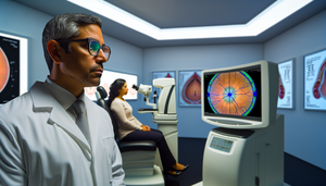
A Hispanic ophthalmologist in his 40s carefully examines the screen of an optical coherence tomography (OCT) machine in a modern clinic. The screen displays a detailed and colorful cross-sectional image of the retina. In the background, a Hispanic female patient in her 30s is seated comfortably. This scene reflects an advanced clinical environment for the diagnosis of eye diseases, particularly retinal conditions.

Advanced Diagnosis of Retinal Diseases in Ophthalmology: The Role of Optical Coherence Tomography

Optical coherence tomography revolutionizes ocular diagnosis with detailed imaging and early detection.

Read more
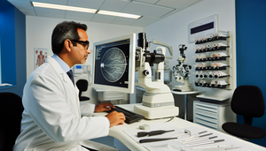
Modern ophthalmology clinic focused on technology. A Hispanic ophthalmologist in his 40s carefully examines a digital retinal scan on a large monitor, highlighting areas of potential retinopathy. The professional and serene environment emphasizes the integration of AI in ophthalmology for retinal analysis and automated retinopathy screening.

AI in Ophthalmology: Automated Systems for Retinal Analysis and Retinopathy Screening

Artificial intelligence is transforming retinal analysis and retinopathy screening, enhancing patient care.

Read more
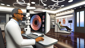
A Hispanic ophthalmologist in his 40s examines a digital retinal image on a large screen in a modern teleophthalmology clinic, while a Hispanic female patient in her 60s waits in the background. This image highlights the advanced technology used in ophthalmic imaging analysis for retinopathy diagnosis, showcasing the role of AI in ophthalmology.

Ophthalmic Imaging Analysis and AI: Advances in Teleophthalmology for Retinopathy Diagnosis

The integration of AI in teleophthalmology enhances the diagnosis of retinopathy and expands visual access.

Read more
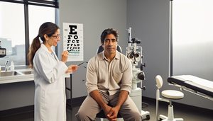
A middle-aged Hispanic man in an ophthalmologist's office, attentively observing a vision chart. The Hispanic female ophthalmologist points to the chart while explaining the significance of early detection and differentiation of retinal dystrophies, such as retinitis pigmentosa, which can lead to night blindness and tunnel vision.

Retinitis Pigmentosa: Early Symptom Identification and Differentiation from Other Retinal Dystrophies

Early detection of retinitis pigmentosa is crucial for accurate differential diagnosis.

Read more
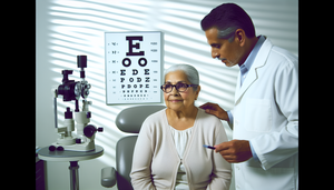
An older Hispanic woman in an ophthalmology consultation, wearing glasses and attentively observing a vision chart. A Hispanic ophthalmologist explains something, pointing to the chart. The scene suggests an eye examination, crucial for the early diagnosis of age-related macular degeneration and differentiation of diabetic retinopathy, utilizing OCT and fluorescein angiography techniques.

Age-Related Macular Degeneration: Early Diagnosis and Differentiation from Diabetic Retinopathy Using OCT and Fluorescein Angiography

Early detection and advanced imaging techniques are crucial for differentiating AMD from DR.

Read more
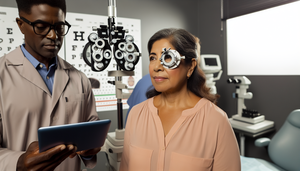
Middle-aged Hispanic woman at an ophthalmological consultation, examining a vision chart. The Hispanic ophthalmologist shows her an eye diagram on a tablet. This professional setting emphasizes the importance of ophthalmological control in patients taking hydroxychloroquine to prevent retinopathy and blurred vision associated with autoimmune diseases.

Hydroxychloroquine: Ophthalmological Monitoring and Key Adverse Effects in Autoimmune Diseases

Hydroxychloroquine, used in autoimmune diseases, necessitates ophthalmological monitoring to prevent irreversible retinopathy.

Read more
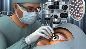
Close-up of a cataract treatment procedure, featuring a Hispanic ophthalmologist performing phacoemulsification surgery. The image shows the surgeon's hands holding a phacoemulsification probe and forceps while making an incision in the cornea of an elderly patient. In the background, modern medical equipment and a monitor displaying an enlarged image of the eye are visible, highlighting the importance of visual recovery and potential surgical complications.

Cataract Treatment: Phacoemulsification Surgery and Intraocular Lens Implant for Optimal Visual Recovery

Phacoemulsification with intraocular lens implantation provides rapid and effective visual recovery.

Read more

Middle-aged Hispanic man consulting with an audiologist while using a hearing aid. The Hispanic audiologist is explaining something by pointing at an ear diagram on a screen. On the desk, there are models of hearing aids and a cochlear implant. The professional and welcoming environment emphasizes the importance of hearing loss treatment, including speech therapy and social adaptation.

Effective Hearing Loss Treatment: Hearing Aids, Cochlear Implants, and Speech Therapy for Social Adaptation

Discover how hearing aids, cochlear implants, and rehabilitation enhance the quality of life in individuals with hearing loss.

Read more
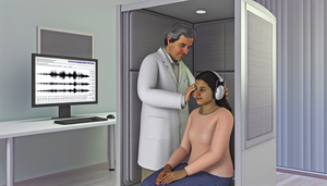
A middle-aged Hispanic audiologist fitting headphones on a young patient in a soundproof booth at an audiology clinic in Spain. In the background, a computer screen displays an audiogram. The image conveys a professional and welcoming environment, perfect for hearing loss diagnosis, including audiometry and otoscopy assessments.

Hearing Loss Diagnosis: Tonal Audiometry, Types, and Reversible Causes

Discover how tonal audiometry aids in identifying hearing loss and its reversible causes.

Read more

A middle-aged Hispanic physician in a white coat discusses a tinnitus treatment plan with a middle-aged Hispanic patient in a modern, serene medical office. The desk features anatomical models of the ear and brochures on tinnitus treatment, emphasizing a multidisciplinary approach that includes TRT, hearing aids, stress management, and experimental drugs.

Tinnitus Treatment: Multidisciplinary Approaches, Including TRT, Hearing Aids, and Stress Management

Discover how retraining therapy and a multidisciplinary approach enhance tinnitus treatment.

Read more
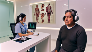
Middle-aged Hispanic man in a modern audiology clinic, wearing headphones connected to an audiometer. A Hispanic audiologist takes notes while conducting an audiometric test for tinnitus diagnosis. On the wall, a diagram of the human ear in English and a digital screen display audiometric test results, highlighting the importance of hearing loss screening and otological causes.

Tinnitus Diagnosis: Audiometry, EMG, and Screening for Otological Causes and Hearing Loss

Audiometry and otoneurological evaluation are essential for an accurate diagnosis of tinnitus.

Read more
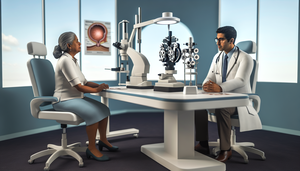
A middle-aged Hispanic ophthalmologist in a white coat and glasses explains retinal detachment treatment options to a Hispanic female patient in her 60s. The table features medical models of the eye illustrating procedures such as pneumatic retinopexy, vitrectomy, and scleral buckle surgery. Advanced ophthalmology equipment is visible in the background. The image emphasizes the educational interaction between the physician and the patient, highlighting the importance of visual success in retinal detachment management.

Effective Treatments for Retinal Detachment: Scleral Buckle, Vitrectomy, and Pneumatic Retinopexy for Optimal Visual Success

Explore surgical options for retinal detachment: pneumatic retinopexy, vitrectomy, and scleral buckle.

Read more
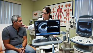
A middle-aged Hispanic patient in an ophthalmologist's office, displaying concern as the Hispanic ophthalmologist explains the results of an ocular examination. The screen shows an OCT scan of the retina with areas of retinal detachment. Nearby, an ultrasound machine suggests its use in diagnosing conditions like torn retina and photopsia. The scene reflects a thorough and attentive medical examination focused on spider vision and patient care.

Retinal Detachment Diagnosis: Symptoms, Photopsia, Torn Retina, OCT, and Ocular Ultrasound

Discover how OCT and ocular ultrasound are essential for diagnosing retinal detachment.

Read more
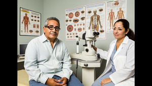
A middle-aged Hispanic man in an ophthalmologist's office, observing an advanced retinal imaging machine. The ophthalmologist, a Hispanic woman, is explaining the procedure with a reassuring smile. On the wall, there are posters about ocular anatomy and diabetic retinopathy, emphasizing the significance of diabetic retinopathy treatment, including panretinal photocoagulation, anti-VEGF therapy, and the necessity of strict glycemic control.

Diabetic Retinopathy Treatment: Laser Photocoagulation, Anti-VEGF, and Strict Glycemic Control

Discover how laser photocoagulation and strict metabolic control are transforming the treatment of diabetic retinopathy.

Read more
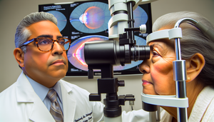
A Hispanic ophthalmologist in his 40s examines the eye of a Hispanic female patient in her 60s using a slit lamp in a modern clinic. In the background, a digital screen displays detailed images of a retina showing signs of diabetic retinopathy, including microaneurysms, hard exudates, neovascularization, and retinal hemorrhages.

Diabetic Retinopathy Diagnosis: Key Findings from Fundoscopy and Fluorescein Angiography

Discover how advanced imaging techniques enhance the early diagnosis of diabetic retinopathy.

Read more
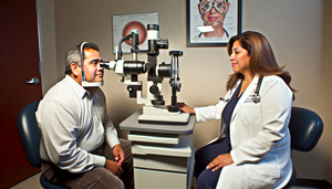
A middle-aged Hispanic patient is seated in front of a slit lamp during an ophthalmology consultation. The doctor, a Hispanic woman in her 40s, is carefully examining the patient's eyes. This image emphasizes the biomicroscopic examination process for cataract diagnosis, highlighting lens opacification and its impact on blurred vision in a professional and trustworthy environment.

Cataract Diagnosis: Biomicroscopic Examination and Refraction for Blurred Vision

Discover how the slit lamp and visual acuity enhance cataract diagnosis.

Read more

In a modern ophthalmology clinic, a middle-aged Hispanic physician discusses glaucoma treatment options with an elderly Hispanic patient. The physician holds a tablet displaying an ocular diagram, while the patient listens attentively. Advanced ophthalmic equipment is visible in the background, highlighting the importance of prostaglandin analogs, topical beta-blockers, laser trabeculoplasty, and filtering surgery in effective glaucoma management.

Glaucoma Treatment: Prostaglandin Analogs, Topical Beta-Blockers, Laser Trabeculoplasty, and Filtering Surgery

Explore personalized strategies for glaucoma treatment, from medications to advanced surgery.

Read more
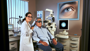
Image of a modern ophthalmology clinic. A middle-aged Hispanic patient is seated in an examination chair while a Hispanic ophthalmologist measures intraocular pressure with a tonometer. In the background, a visual field analyzer and a 3D image of the optic nerve on a monitor emphasize the significance of these tests in glaucoma diagnosis, including the assessment of optic nerve cupping and the distinction between open angle vs closed angle glaucoma.

Glaucoma Diagnosis: Intraocular Pressure, Gonioscopy, and Assessing Optic Nerve Cupping in Open Angle vs Closed Angle

Discover how tonometry, perimetry, and optic nerve assessment are crucial for the early diagnosis of glaucoma.

Read more
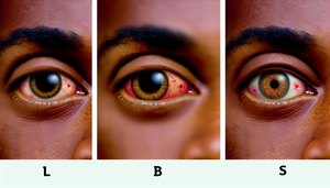
Close-up of three human eyes displaying different types of conjunctivitis: the left eye with bacterial conjunctivitis shows conjunctival hyperemia and yellowish ocular discharge; the central eye with viral conjunctivitis exhibits conjunctival hyperemia and watery ocular discharge; the right eye with allergic conjunctivitis presents conjunctival hyperemia, swelling, and watery ocular discharge.

Conjunctivitis Diagnosis: Differentiating Bacterial, Viral, and Allergic Causes with Ocular Discharge and Conjunctival Hyperemia

Discover how to differentiate between bacterial, viral, and allergic conjunctivitis for effective treatment.

Read more

A Hispanic physician in his 40s, wearing a white coat and stethoscope, examines the ear of an 8-year-old Hispanic boy with an otoscope in a medical examination room. The child appears calm, while the physician demonstrates a professional and careful demeanor. In the background, a medical poster illustrates ear anatomy, emphasizing the significance of accurate middle ear diagnosis, particularly in identifying a bulging tympanic membrane associated with bacterial infections and ear pain.

Middle Ear Diagnosis: Otoscopy for Bulging Tympanic Membrane and Bacterial Infection in Ear Pain

Discover how otoscopy aids in differentiating between otitis media and externa for precise treatment.

Read more

Modern medical consultation with a Hispanic doctor explaining sinusitis treatment options to a patient. The doctor holds a tablet displaying a 3D model of the paranasal sinuses. On the desk, there are brochures about antibiotics, decongestants, and endoscopic sinus surgery. The room is decorated with anatomical posters of the head and paranasal sinuses, creating a professional and collaborative environment.

Effective Sinusitis Treatment: Antibiotics, Decongestants, and Endoscopic Surgery Options for Physicians

Explore the comprehensive management of sinusitis with antibiotics, decongestants, and endoscopic sinus surgery.

Read more
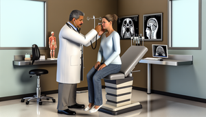
A middle-aged Hispanic physician examines a Hispanic patient in a modern ENT examination room. He uses an otoscope to perform a rhinoscopy, assessing the patient's paranasal sinuses for signs of bacterial sinusitis. In the background, a CT scan of the paranasal sinuses is displayed on a computer screen alongside an anatomical model of the human head. The room is well-lit and organized, creating a professional and welcoming environment for accurate sinusitis diagnosis.

Sinusitis Diagnosis: ENT Examination and Imaging Tests for Bacterial Sinusitis

Discover how rhinoscopy and sinus CT enhance the diagnosis of sinusitis.

Read more