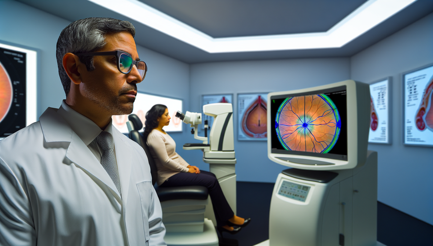
Advanced Diagnosis of Retinal Diseases in Ophthalmology: The Role of Optical Coherence Tomography

The optical coherence tomography (OCT) has revolutionized the field of ophthalmology by providing detailed and non-invasive images of the retina. This technology has enabled significant advancements in the diagnosis of eye diseases, particularly those affecting the retina. Since its introduction, OCT has evolved to offer high-resolution images that approach histological levels, allowing ophthalmologists to detect and monitor a variety of retinal pathologies with unprecedented accuracy.
Diving Deeper into Optical Coherence Tomography
OCT is based on light backscattering to create detailed images of ocular structures. One of the most recent innovations is optical coherence tomography angiography (OCTA), which allows visualization of the microvasculature of the retina and choroid without the need for dyes. This technique has proven particularly useful in the diagnosis of vascular diseases of the retina, such as diabetic retinopathy and age-related macular degeneration.
Furthermore, OCT has been fundamental in understanding neuro-ophthalmological diseases. Recent studies have shown that OCT can detect structural and functional changes in the early stages of diseases such as multiple sclerosis and Alzheimer’s, even before evident clinical symptoms appear (see study). This early detection capability is crucial for the timely management and treatment of these conditions.
OCT has also been used to identify and characterize less common conditions such as acute paracentral maculopathy, providing a deeper understanding of its pathophysiology and guiding future research.
Conclusions
Optical coherence tomography has transformed the diagnosis and management of retinal diseases. Its ability to provide detailed and non-invasive images has significantly improved our understanding of ocular pathophysiology and has enabled more accurate and early diagnosis of various pathologies. As technology continues to advance, it is likely that OCT will continue to play a crucial role in ophthalmology, enhancing clinical outcomes and the quality of life for patients.
References
- [1] Optical coherence tomography angiography: A comprehensive review of current methods and clinical applications
- [2] Optical coherence tomography angiography in neuro-ophthalmology
- [3] Paracentral acute middle maculopathy-review of the literature
Created 24/1/2025
