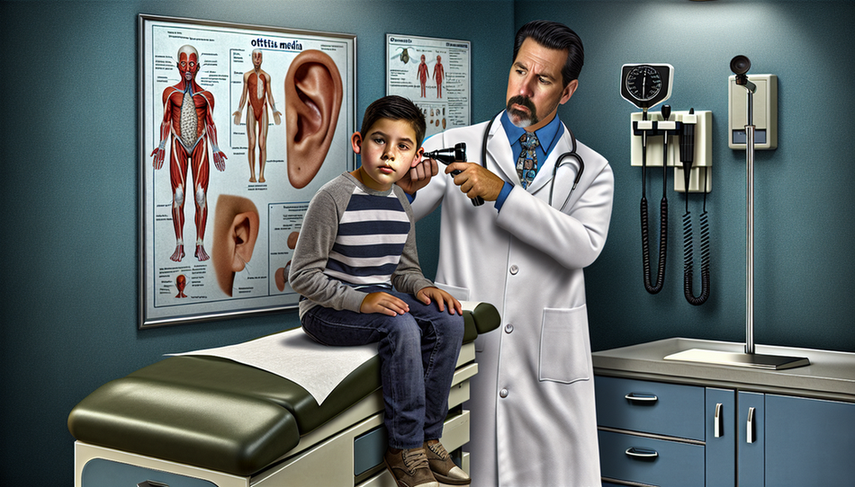
Middle Ear Diagnosis: Otoscopy for Bulging Tympanic Membrane and Bacterial Infection in Ear Pain

Dear colleagues, otitis media is one of the most common infections in pediatric practice, and accurate diagnosis is crucial for effective treatment. Otoscopy is an essential tool in this process, allowing us to visualize the bulging tympanic membrane and other characteristic signs of this condition. However, it is equally important to differentiate it from otitis externa, as both conditions require distinct therapeutic approaches.
Diving Deeper into Diagnosis
Otitis media is characterized by the presence of fluid in the middle ear, accompanied by symptoms such as ear pain and irritability in younger patients. Otoscopy allows us to observe signs such as the bulging tympanic membrane, which may appear opaque or have reduced mobility, indicating a bacterial infection in the middle ear. In contrast, otitis externa affects the external auditory canal and presents with inflammation, pain upon touch, and often purulent discharge.
A study on otoscopic examination in horses highlights the importance of proper visualization of the auditory canal and tympanic membrane, which is equally applicable in humans for accurate diagnosis. Additionally, otitis externa in animals is described as a syndrome reflecting underlying dermatological diseases, underscoring the need for a systematic diagnostic approach that includes clinical history, otoscopy, and cytology.
Conclusions
The differentiation between otitis media and externa is fundamental to avoid inappropriate treatments and complications. Otoscopy remains an invaluable tool in this process, allowing us to identify key features such as the bulging tympanic membrane and inflammation of the auditory canal. By integrating these findings with clinical history and other complementary examinations, we can ensure accurate diagnosis and appropriate management of these common yet potentially complicated conditions.
Referencias
- [1] Otoscopic, cytological, and microbiological examination of the equine external ear canal.
- [2] Diagnosis and medical treatment of otitis externa in the dog and cat.
Created 5/1/2025
