Diabetic Retinopathy Diagnosis: Key Findings from Fundoscopy and Fluorescein Angiography
Diabetic retinopathy is one of the most common microvascular complications of diabetes and a leading cause of blindness in working-age adults. Early and accurate diagnosis of diabetic retinopathy is crucial to prevent disease progression and preserve vision. In this context, fundoscopy and fluorescein angiography are essential diagnostic tools that allow for detailed evaluation of the retinal vasculature and identification of characteristic signs such as microaneurysms, hard exudates, neovascularization, and retinal hemorrhages.
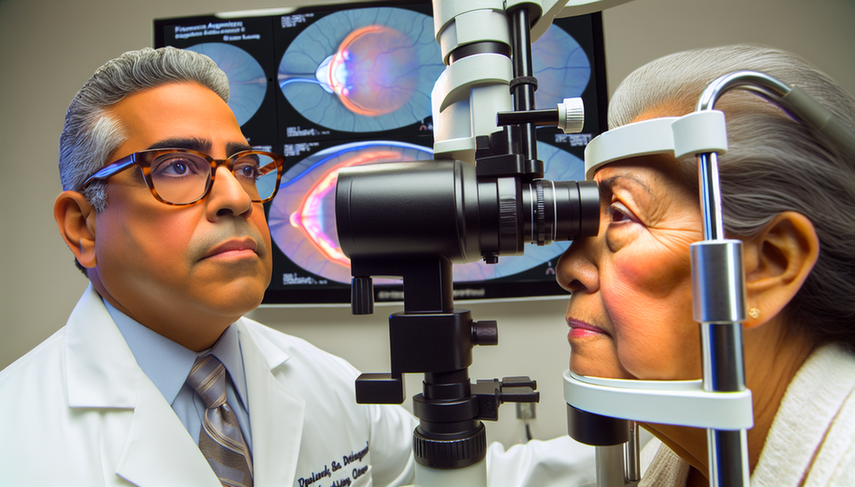
Fundoscopy is a non-invasive technique that enables direct visualization of the retina and its vascular structures. Through this technique, early changes in the retina, such as microaneurysms and hard exudates, which are indicative of diabetic retinopathy, can be identified. Fluorescein angiography, on the other hand, is a procedure that uses a fluorescent dye to highlight blood vessels in the retina, allowing for a more detailed assessment of perfusion and the presence of neovascularization or retinal hemorrhages. This technique has been fundamental since its introduction in 1961, providing a clearer view of vascular alterations in diabetic retinopathy [1].
In recent years, the development of new imaging technologies, such as optical coherence tomography (OCT) and OCT angiography (OCT-A), has revolutionized clinicians' ability to examine the retinal microvasculature without the need for contrast injections. These techniques allow for three-dimensional and in-depth visualization of the retinal vascular structure, improving the accuracy in detecting microvascular changes and capillary perfusion loss [2]. Additionally, adaptive optics has enabled the study of microscopic features of the vascular and neuronal tissues of the retina, providing new insights into the development and treatment response of diabetic retinopathy [3].
In conclusion, the diagnosis of diabetic retinopathy has greatly benefited from advances in imaging techniques. Fundoscopy and fluorescein angiography remain fundamental tools, but new technologies such as OCT and OCT-A are enhancing diagnostic accuracy and allowing for a more detailed evaluation of the retinal microvasculature. These advancements not only facilitate early detection of the disease but also provide a better understanding of its progression and treatment response, which is crucial for improving visual outcomes in patients with diabetes.
Referencias
- [1] New developments in angiography for the diagnosis and management of diabetic retinopathy.
- [2] Imaging in diabetic retinopathy.
- [3] Adaptive Optics in the Evaluation of Diabetic Retinopathy.
Created 6/1/2025