Retinal Detachment Diagnosis: Symptoms, Photopsia, Torn Retina, OCT, and Ocular Ultrasound
The retinal detachment is an ophthalmological emergency that requires rapid and accurate diagnosis to prevent permanent vision loss. Initial symptoms may include photopsia (flashes of light), spider vision or shadows in the visual field, and the perception of a curtain falling over vision. These symptoms are indicative of a possible torn retina, which can lead to complete detachment if not treated appropriately.
Diving Deeper into Diagnosis
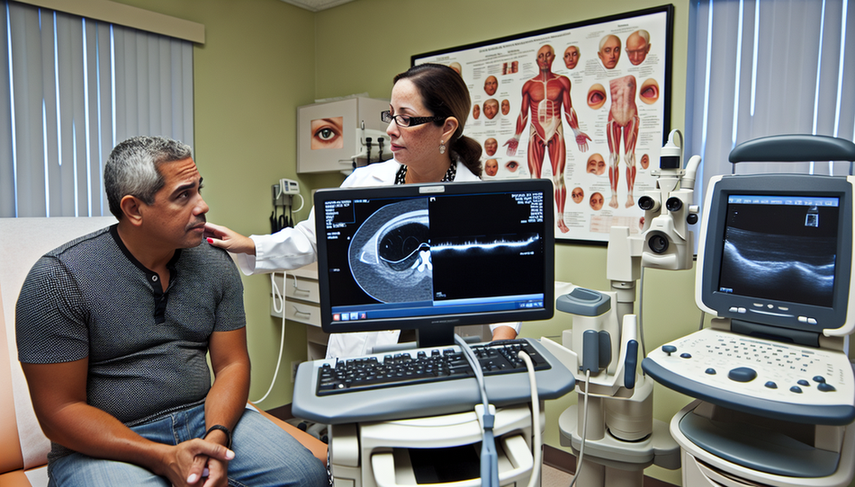
The diagnosis of retinal detachment relies on a combination of advanced imaging techniques. Optical coherence tomography (OCT) is a crucial tool that provides high-resolution images of the retinal layers, facilitating the identification of tears and detachments. A recent study has shown that OCT is particularly useful for detecting structural changes in the retina, such as retinal displacement, which can be a sign of detachment [1].
Additionally, ocular ultrasound is a complementary technique used to assess the status of the vitreous and retina, especially in cases where opacities in the ocular media prevent clear visualization through other methods. Ultrasound is effective in confirming the presence of retinal detachment and evaluating its extent [2].
In the context of pediatric vitreoretinal surgery, intraoperative OCT has proven to be a valuable tool for enhancing surgical safety and efficiency, allowing for the identification of subtle retinal tears and other findings that may contribute to the development of retinal detachments [3].
Conclusions
Early and accurate diagnosis of retinal detachment is essential for preserving patient vision. The combination of imaging techniques such as OCT and ocular ultrasound provides a detailed evaluation of the retina, enabling ophthalmologists to make informed treatment decisions. The integration of these technologies into daily clinical practice significantly improves visual outcomes for patients at risk of retinal detachment.
Referencias
- [1] Optical coherence tomography homography for detection of retinal displacement: a validation study.
- [2] Optical coherence tomography for diagnosis of posterior vitreous detachment at the macular region.
- [3] Pediatric Vitreoretinal Surgery and Integrated Intraoperative Optical Coherence Tomography.
Created 6/1/2025