
Neurology

Navigate the existing neurology articles covering diagnosis, management and neurological research.
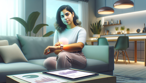
Young Hispanic woman sitting on a modern sofa, wearing a smartwatch that monitors her health. On the table, a tablet displays graphs and notifications related to seizure detection and remote monitoring of epileptic episodes. The scene illustrates the integration of modern technology in daily life, emphasizing the importance of alert devices for medical professionals.

Wearables for Seizure Detection: Early Alert Devices and Remote Monitoring of Epileptic Episodes

Wearables enhance the detection and remote management of seizures, enabling faster and more effective interventions.

Read more
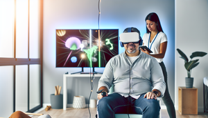
A middle-aged Hispanic man engages in cognitive exercises using a virtual reality headset in a modern neurological rehabilitation clinic. A young Hispanic therapist observes him while monitoring his progress on a tablet. The bright and serene room enhances the therapeutic experience.

Neurological Rehabilitation with Virtual Reality: Interactive Cognitive Exercises and Functional Diagnosis Techniques

Virtual reality is revolutionizing neurological rehabilitation with personalized cognitive exercises and advanced functional diagnostics.

Read more
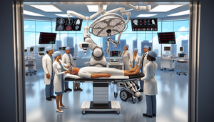
A modern operating room equipped with advanced technology where a robotic neurosurgery system performs brain surgery on a middle-aged Hispanic patient. A diverse team of surgeons observes and controls the procedure through a digital interface. Nearby, a young Hispanic woman uses an exoskeleton, guided by a physical therapist, to assist her in walking. The scene embodies innovation and hope in medical science, highlighting the integration of robotic neurosurgery and guided rehabilitation.

Robotic Neurosurgery and Exoskeletons: A New Frontier in Brain Surgeries and Guided Rehabilitation

Robotic neurosurgery and exoskeletons are transforming surgical precision and neurological rehabilitation.

Read more
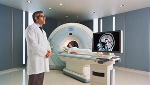
Modern medical imaging room featuring an advanced functional MRI. A Hispanic neurologist in his 40s examines a digital brain map on a high-resolution monitor, displaying brain activity across various regions. In the background, a Hispanic female patient in her 30s is calmly positioned inside the MRI machine. The image emphasizes advanced technology and precision in early neurological diagnosis and lesion detection.

Functional MRI and Brain Mapping: Key Tools for Early Neurological Diagnosis and Lesion Detection

Functional MRI and brain mapping are revolutionizing early neurological diagnosis by detecting alterations before evident structural damage occurs.

Read more
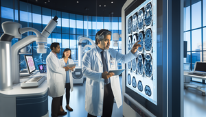
A Hispanic neurologist in his 40s carefully analyzes digital brain scans on a large screen in a modern neurology lab. In the background, a diverse team of medical professionals discusses brain images on a tablet. The scene emphasizes the role of AI in neurology, particularly in neuroimaging analysis for studying neurodegenerative diseases.

AI in Neurology: Intelligent Neuroimaging Analysis for Neurodegenerative Diseases Using Magnetic Resonance Imaging

AI enhances the early diagnosis of neurodegenerative diseases through advanced neuroimaging analysis.

Read more
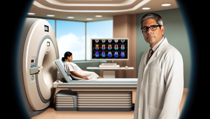
Modern hospital room featuring a middle-aged Hispanic physician analyzing brain scans on a digital screen, showcasing the innovative use of functional magnetic resonance imaging in the diagnosis of neurological disorders. In the background, a Hispanic patient is preparing for her scan in an advanced MRI machine.

Neuroimaging and Functional Magnetic Resonance Imaging: Innovations in the Diagnosis of Neurological Disorders

Discover how functional magnetic resonance imaging is revolutionizing the diagnosis of neurological diseases.

Read more
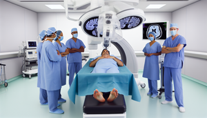
Modern neurosurgery operating room featuring advanced technology, highlighting a surgical robotic arm over a middle-aged Hispanic patient. A diverse team of neurosurgeons, including a Hispanic woman and an Asian man, monitors the procedure. A digital screen displays a 3D brain scan, emphasizing the operative precision of the surgical navigation system and the role of AI in neurosurgery.

AI in Neurosurgery: Enhancing Operative Precision with Robotics and Surgical Navigation

Robotics and AI are revolutionizing neurosurgery with enhanced precision and safety.

Read more
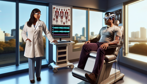
Modern medical examination room focused on neuromuscular diseases diagnosis. A Hispanic male patient in his 40s is connected to an advanced electromyography (EMG) machine with digital screens displaying graphs and data. A Hispanic female doctor in her 50s analyzes the results on a tablet, emphasizing the role of smart electromyography and artificial intelligence in rapid diagnosis and neurological prognosis.

Differential Diagnosis of Neuromuscular Diseases Using Smart Electromyography: Rapid Signal Classification for Enhanced Neurological Prognosis

Smart electromyography with AI enhances the rapid and accurate diagnosis of neuromuscular diseases.

Read more
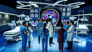
Diverse medical team of Hispanic doctors and nurses in a modern hospital, analyzing a digital screen displaying a brain image. The screen highlights neural pathways, indicating the use of diagnostic algorithms and guided thrombolysis for effective stroke management. The environment reflects innovation and collaboration in immediate neurological care.

Stroke Management with AI: Diagnostic Algorithms and Guided Thrombolysis for Immediate Neurological Care

Artificial intelligence is revolutionizing stroke management by enhancing diagnosis and personalizing treatment.

Read more
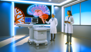
A Hispanic male physician in his 40s, wearing a white coat and glasses, points to a computer screen displaying a 3D brain MRI with highlighted neural networks. Next to him, a Hispanic female physician in her 30s reviews data on a digital tablet. The scene illustrates the use of advanced technology, including deep learning, in the diagnosis of neurological disorders.

Neural Networks and Neuroimaging: AI in the Diagnosis of Brain MRI and Neurological Disorders

Artificial intelligence and neural networks enhance the accuracy and speed of neurological diagnosis.

Read more
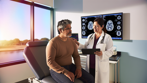
A middle-aged Hispanic patient is seated at an examination table in a modern medical room. A Hispanic female doctor, dressed in a white coat, is showing a brain scan on a tablet, highlighting relevant areas. On the wall, a large screen displays a 3D model of a human brain with highlighted neural pathways, symbolizing the use of AI in Parkinson's diagnosis. The atmosphere is professional and reassuring, reflecting advancements in clinical algorithms and neurological telemedicine for monitoring movement disorders.

AI-Assisted Parkinson's Diagnosis: Enhancing Clinical Algorithms and Monitoring Systems in Neurological Telemedicine

AI is revolutionizing Parkinson's diagnosis, enhancing accuracy and facilitating neurological telemedicine.

Read more
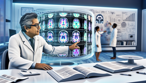
A middle-aged Hispanic scientist examines a high-resolution digital brain scan on a monitor in a medical research laboratory. The scan displays vibrant colors highlighting different brain regions, symbolizing advanced imaging techniques. In the background, a diverse group of researchers discusses in front of a whiteboard filled with diagrams and notes, reflecting collaboration in advanced Alzheimer research, focusing on early detection, cognitive assessment, and the role of AI in neurology.

AI in Early Detection of Alzheimer’s: New Digital Biomarkers and Cognitive Assessment Techniques

Artificial intelligence enhances early detection of Alzheimer's through digital biomarkers and cognitive analysis.

Read more
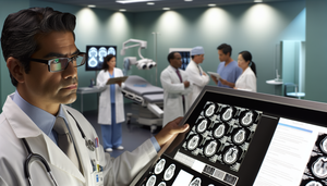
Image of a Hispanic neurosurgeon in his 40s reviewing brain scans of a meningioma in a modern hospital. He is wearing a white coat and glasses, displaying a focused expression. In the background, a diverse medical team discusses with charts and tablets, reflecting a collaborative environment in a high-tech setting, emphasizing meningioma prognosis, life expectancy, and survival factors.

Meningioma Prognosis and Life Expectancy: Key Prognostic Factors for Neurosurgeons

Total resection and molecular characterization improve prognosis and survival in meningiomas.

Read more
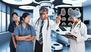
Diverse team of healthcare professionals in a modern hospital discussing a brain scan on a digital screen. A middle-aged Hispanic neurologist points to the scan while explaining to a young Asian nurse taking notes on a tablet and an older Caucasian radiologist listening attentively. The setting emphasizes a multidisciplinary approach to stroke care, highlighting warning signs, acute management, and rehabilitation strategies.

Essential Neurology: Recognizing Stroke Warning Signs and Multidisciplinary Acute Management

Discover how a multidisciplinary approach optimizes stroke management and enhances recovery.

Read more
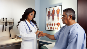
A middle-aged Hispanic patient, dressed in a light blue hospital gown, is seated at an examination table in a modern, well-lit medical room. A Hispanic doctor, wearing a white coat and stethoscope, gently holds the patient's hand while assessing his reflexes with a reflex hammer. The doctor's expression is attentive and focused, while the patient appears calm and cooperative. In the background, anatomical charts of the brain and nervous system are visible, emphasizing the importance of thorough neurological evaluations for the early diagnosis of neurodegenerative diseases.

Neurological Evaluations: Recognizing Subtle Signs for Early Diagnosis of Neurodegenerative Diseases

Discover how to identify subtle signs for the early diagnosis of neurodegenerative diseases.

Read more
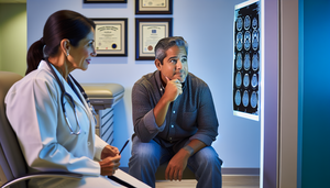
Middle-aged Hispanic patient in a medical consultation, displaying concern as the Hispanic doctor explains a contrast-enhanced MRI of the brain on a lightbox. The doctor points to a specific area in the image, emphasizing the importance of MRI in diagnosing astrocytomas and assessing focal symptoms. The setting is professional and calm, highlighting the significance of medical consultations and advanced imaging techniques in the evaluation of primary brain tumors.

Astrocytoma: Recognizing Focal Symptoms and the Importance of Contrast-Enhanced MRI in Primary Brain Tumors

Early detection of focal symptoms and the use of contrast-enhanced MRI improve the diagnosis of astrocytomas.

Read more
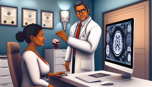
A middle-aged Hispanic woman attentively examines a computer screen displaying a detailed brain scan in a doctor's office. A Hispanic doctor in his 50s, wearing a white coat and glasses, points to the image with a pen while explaining the scan. This scene emphasizes the critical role of neuroimaging in the early detection of meningiomas, particularly in patients presenting with morning headaches and personality changes.

Meningioma: Morning Headaches, Personality Changes, and the Role of Neuroimaging in Early Detection

Early detection of meningiomas through neuroimaging enhances clinical management despite nonspecific symptoms.

Read more
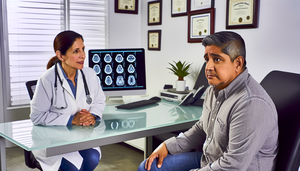
A middle-aged Hispanic patient with a concerned expression attentively listens to a Hispanic doctor explaining the results of a magnetic resonance imaging (MRI) scan. The image on the screen highlights areas suggesting a possible diagnosis of glioblastoma. This interaction underscores the importance of communication in the early diagnosis of neurological symptoms such as persistent headache and epileptic seizures.

Early Neurological Symptoms of Glioblastoma: The Importance of MRI for Timely Diagnosis of Persistent Headaches and Epileptic Seizures

Magnetic resonance imaging is crucial for the early diagnosis of glioblastoma, enhancing prognosis.

Read more
A young Hispanic girl, approximately 3 years old, sitting on a colorful play mat in a pediatric clinic. She is wearing a light pink dress and holding a stuffed toy. In the background, a warm-smiling Hispanic pediatrician observes. The image reflects a caring environment, relevant for the early detection of Rett syndrome, developmental regression, and differentiation from autism spectrum disorder, emphasizing the importance of the MECP2 genetic test.

Rett Syndrome: Key Insights for Early Detection and Differentiation from Autism Spectrum Disorder

Discover how to differentiate Rett syndrome from autism through clinical signs and genetic testing.

Read more

A Hispanic boy approximately 10 years old sitting at a school desk, displaying an involuntary facial expression indicative of a transient tic. The scene takes place in a bright and welcoming classroom, with other children focused on their tasks and a Hispanic teacher explaining mathematical equations on the blackboard, highlighting the context of Tourette syndrome and the differentiation of motor and vocal tics.

Tourette Syndrome: Recognizing Persistent Motor and Vocal Tics and Differentiating Them from Transient Tics in ADHD Comorbidity

Discover how to differentiate between transient and persistent tics in Tourette syndrome for effective management.

Read more

Middle-aged Hispanic patient in a modern medical consultation, attentively listening to a Hispanic doctor explaining the diagnosis of Fabry disease using a digital tablet. This interaction emphasizes effective communication in diagnosing Fabry disease, differentiating it from neurological conditions such as multiple sclerosis, while also addressing issues like neuropathic pain and corneal opacity related to alpha-galactosidase deficiency.

Fabry Disease: Diagnostic Keys and Differentiation of Neurological Processes in Alpha-Galactosidase Deficiency and Multiple Sclerosis

Discover how to differentiate Fabry disease from multiple sclerosis using essential diagnostic clues.

Read more
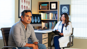
A middle-aged Hispanic man in a modern medical office, looking thoughtful, facing a Hispanic female doctor who is listening attentively. On the desk, a screen displays a brain scan image, indicating a discussion about Creutzfeldt-Jakob disease, rapidly progressive dementia, and the differentiation of encephalopathies, including the role of the 14-3-3 protein and EEG findings.

Creutzfeldt-Jakob Disease: Recognizing Rapidly Progressive Dementia and Differentiating from Other Encephalopathies Using 14-3-3 Protein and EEG

Discover how to differentiate Creutzfeldt-Jakob disease from other treatable encephalopathies using key markers.

Read more
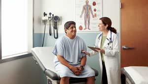
Middle-aged Hispanic patient in a modern medical examination room, attentively listening to a Hispanic doctor who is explaining with a reassuring smile. The image conveys a professional and warm environment, ideal for discussing topics such as Shy-Drager syndrome, autonomic dysfunction, and atypical parkinsonism, with an emphasis on the importance of magnetic resonance imaging in differential diagnosis.

Shy-Drager Syndrome: Assessing Autonomic Dysfunction and Differential Diagnosis of Atypical Parkinsonism Using Magnetic Resonance Imaging

Discover how MRI and autonomic tests differentiate Shy-Drager syndrome from other parkinsonisms.

Read more
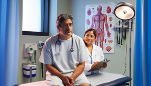
A middle-aged Hispanic patient with an attentive and slightly worried expression is seated on an examination table in a medical office. A Hispanic female doctor, wearing a white coat and stethoscope, is reassuringly explaining something while holding a clipboard. Behind them, there is an anatomical chart of the human nervous system. The scene reflects a professional and caring environment, pertinent to the discussion on Guillain-Barré syndrome, ascending weakness, EMG findings, and the differentiation from chronic polyradiculoneuropathy, including lumbar puncture considerations.

Early Detection of Guillain-Barré Syndrome: Differentiating from Chronic Polyradiculoneuropathy Using EMG and Lumbar Puncture

Early detection and differentiation between GBS and CIDP is crucial for optimal treatment.

Read more
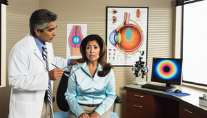
A middle-aged Hispanic woman in an ophthalmologist's office, displaying concern as the Hispanic doctor explains something while pointing to an anatomical chart of the eye highlighting the optic nerve. On the desk, there are medical tools and a computer with a 3D model of the eye. The scene reflects a professional and modern environment, relevant for discussions on optic neuritis, sudden vision loss, and multiple sclerosis differentiation, including the evoked potential test.

Optic Neuritis: Key Symptoms, Risk Assessment for Multiple Sclerosis, and the Role of Evoked Potential Tests in Sudden Vision Loss and Eye Pain

Early identification of optic neuritis is crucial for assessing the risk of multiple sclerosis.

Read more
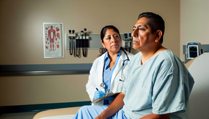
A middle-aged Hispanic patient in a modern medical examination room, attentively listening to a Hispanic doctor explaining multisystem atrophy, dysautonomia, and the differentiation of parkinsonism from other parkinson plus syndromes using magnetic resonance imaging.

Multisystem Atrophy: Recognizing and Differentiating It from Other Parkinson Plus Syndromes Using MRI and Dysautonomia Insights

Discover how to differentiate multiple system atrophy from other Parkinson-plus syndromes using advanced techniques.

Read more
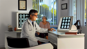
Brightly lit medical office where a middle-aged Hispanic doctor carefully examines a brain PET scan on a lightbox, highlighting areas associated with frontotemporal dementia. In the background, an older Hispanic woman observes the doctor. The scene reflects a professional and welcoming environment, emphasizing the importance of early diagnosis and differentiation from Alzheimer's disease, particularly in cases involving behavioral changes and progressive aphasia.

Frontotemporal Dementia: Early Diagnosis and Differentiation from Alzheimer's Disease through Behavioral Changes and Brain PET Imaging

Early differentiation between frontotemporal dementia and Alzheimer's enhances clinical management through neuroimaging and biomarkers.

Read more
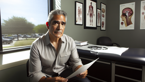
A middle-aged Hispanic man in a modern medical office, focused on a medical chart. Next to him are medical books and a stethoscope, with anatomical charts on the wall, prominently featuring one of the brain. The scene reflects a medical research environment related to corticobasal degeneration, asymmetric rigidity, apraxia, and the differentiation of atypical parkinsonism through magnetic resonance imaging.

Corticobasal Degeneration: Recognizing Asymmetric Rigidity and Apraxia for Differentiation from Atypical Parkinsonism Using Magnetic Resonance Imaging

Discover how to differentiate corticobasal degeneration from other atypical parkinsonisms through clinical evaluation and magnetic resonance imaging.

Read more
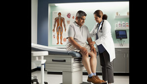
A middle-aged Hispanic patient sitting on an examination table in a modern medical office. A Caucasian doctor is carefully examining the patient's foot, illustrating a clinical evaluation of peripheral neuropathy. The wall features a diagram of the human nervous system, highlighting the importance of sensitivity testing and conduction velocity in the differentiation of diabetic polyneuropathy, with a focus on distal tingling.

Peripheral Neuropathy: Clinical Evaluation and Differentiation of Diabetic Polyneuropathy through Sensitivity Testing and Conduction Velocity Analysis

Discover how to differentiate diabetic polyneuropathy through effective clinical evaluation and sensitivity testing.

Read more
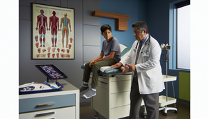
Medical consultation in a modern clinic: a Hispanic physician in his 40s examines the foot of a Hispanic teenager, pointing to a diagram of the nervous system on a tablet. On the wall, a poster about the anatomy of peripheral nerves. This scene emphasizes the importance of early diagnosis of Charcot-Marie-Tooth disease, hereditary peripheral neuropathy, and the role of electromyography in assessing foot deformities and differentiating ataxias.

Charcot-Marie-Tooth Disease: Early Diagnosis and Evaluation of Hereditary Peripheral Neuropathy through Electromyography and Differentiation of Ataxias

Early diagnosis of Charcot-Marie-Tooth disease enhances patient management and quality of life.

Read more
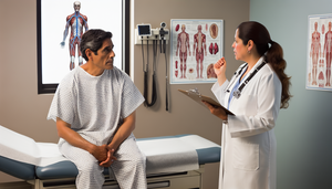
A middle-aged Hispanic patient with a concerned expression is sitting on an examination table while a Hispanic doctor, wearing a white coat and stethoscope, reassuringly explains something to him. She holds a clipboard, and on the wall, there is an anatomical chart of the nervous system, emphasizing the importance of early detection and differential diagnosis in neuromuscular disorders such as myasthenia gravis and Lambert-Eaton syndrome, including the role of the edrophonium test and anti-AChR antibodies in assessing fluctuating muscle weakness.

Myasthenia Gravis: Early Detection and Differentiation from Lambert-Eaton Syndrome Using Edrophonium Test and Anti-AChR Antibodies

Discover how to differentiate myasthenia gravis from Lambert-Eaton syndrome for accurate diagnosis.

Read more
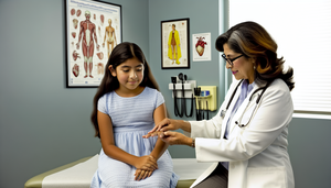
A Hispanic girl approximately 10 years old is sitting in a consultation room with a middle-aged Hispanic female doctor, who is carefully examining the girl's hand. The scene reflects a professional and caring environment, relevant for the diagnosis of Sydenham's chorea, differentiation from other causes of involuntary movements, and the assessment of rheumatic fever through the ASO test.

Sydenham's Chorea: Diagnosis and Differentiation from Involuntary Movements and Huntington's Disease in Rheumatic Fever

Discover how to diagnose Sydenham's chorea and differentiate it from other causes of involuntary movements.

Read more
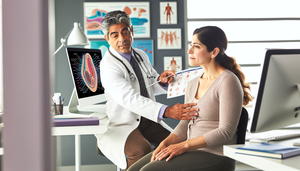
A middle-aged Hispanic woman reviews a medical chart in a modern office while a Hispanic doctor explains something, pointing to a diagram of the nervous system on a screen. The scene reflects clear communication and support, ideal for a blog focused on multiple sclerosis diagnosis, initial symptoms, brain MRI findings, and differentiation from neuromyelitis optica.

Multiple Sclerosis Diagnosis: Recognizing Initial Symptoms and Differentiating from Neuromyelitis Optica Using Brain MRI and Oligoclonal Bands

Discover how to differentiate multiple sclerosis from neuromyelitis optica using MRI and biomarkers.

Read more
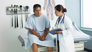
Middle-aged Hispanic patient in a modern medical examination room, wearing a light blue hospital gown. A Caucasian doctor in a white coat and stethoscope attentively listens to the patient while holding a clipboard. A diagram of the human nervous system is visible on the wall. The professional and organized environment emphasizes clinical assessment in the context of ALS evaluation, including electromyography and nerve conduction studies for neuropathy differentiation.

ALS Assessment: Clinical Evaluation and Differentiation of Neuropathies through Electromyography and Nerve Conduction Studies

Discover how clinical evaluation and advanced biomarkers enhance the differential diagnosis of ALS.

Read more
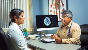
A middle-aged Hispanic man in a modern medical consultation, attentively listening to a Hispanic female doctor holding a clipboard and wearing a stethoscope. On the desk, a computer displays a brain scan image, and a poster of the nervous system is on the wall. The scene reflects a professional and calm medical consultation focused on the early diagnosis of Parkinson's, essential tremor, muscle rigidity, and the differentiation of parkinsonism using the DaTscan test.

Early Diagnosis of Parkinson's: Key Insights for Differentiating Parkinsonism and Essential Tremor with DaTscan

Discover how biomarkers and advanced neuroimaging are revolutionizing the early diagnosis of Parkinson's disease.

Read more

Bright medical office illuminated by natural light, where a middle-aged Hispanic doctor in a white coat and glasses carefully examines an image of a brain on a computer screen. The image highlights specific areas of the brain, indicating a focus on the early diagnosis of Alzheimer’s, differentiation of vascular dementia, and the use of biomarkers. In the background, a bookshelf filled with medical books and a plant create a warm and professional atmosphere.

Early Diagnosis of Alzheimer’s: Differentiating from Vascular Dementia and the Role of Biomarkers in Mild Cognitive Impairment

Discover how biomarkers and neuroimaging enhance early diagnosis and differentiation of Alzheimer's disease.

Read more
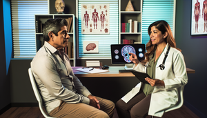
A middle-aged Hispanic patient in a modern medical consultation, attentively listening to a Hispanic doctor explaining the treatment of partial seizures with levetiracetam using a brain diagram on a tablet. The scene reflects a professional and trustworthy consultation focused on discussing dose adjustment and the management of irritability and fatigue associated with antiepileptics.

Levetiracetam: Behavioral Side Effects, Dose Adjustment, and Management of Irritability and Fatigue in Partial Seizures

Explore the behavioral effects of levetiracetam and how to adjust its dosage in epilepsy.

Read more

A middle-aged Hispanic physician with a thoughtful expression sits at a round table in a naturally lit hospital room. Dressed in a white coat and stethoscope, he has open medical books and handwritten notes in front of him. An older Hispanic woman, with a calm smile, listens attentively. The scene conveys warmth, professionalism, and empathy, creating a trusting environment to discuss topics such as dementia, delirium, cognitive impairment, and attentional fluctuations, emphasizing the importance of neuropsychological evaluation.

Dementia vs. Delirium: Guidelines for Differentiating Cognitive Impairment and Attentional Fluctuations through Neuropsychological Evaluation

Discover how to differentiate between dementia and delirium to enhance patient treatment and outcomes.

Read more
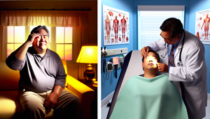
Split image depicting two scenarios: on the left, a middle-aged Hispanic man in a living room, showing signs of discomfort indicative of cluster headache; on the right, a young Hispanic woman in a clinical setting, being examined by a healthcare professional, focusing on the jaw and temple area, suggesting trigeminal neuralgia and intense facial pain.

Cluster Headache vs. Trigeminal Neuralgia: Key Clinical Insights for Managing Intense Facial Pain and Triggers

Discover the key clinical differences between cluster headaches and trigeminal neuralgia for effective management.

Read more
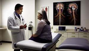
A middle-aged Hispanic physician in a white coat and stethoscope explains to a Hispanic female patient in her 30s, pointing to an anatomical chart of the nervous system on the wall. The patient, seated on an examination table, listens attentively. The well-lit and equipped medical consultation room emphasizes the importance of understanding demyelinating disorders such as Guillain-Barré syndrome and chronic demyelinating polyneuropathy, as well as the role of lumbar puncture and electromyography in diagnosis.

Guillain-Barré Syndrome vs. CIDP: How to Differentiate Demyelinating Disorders in Ascending Weakness

Discover how to differentiate Guillain-Barré syndrome from chronic demyelinating polyneuropathy for effective treatment.

Read more
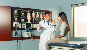
A middle-aged Hispanic doctor in a white coat and stethoscope gestures while explaining to a young Hispanic patient sitting on an examination table in a well-lit office. On the wall, there is a medical chart featuring diagrams of the brain and heart, indicating topics related to epileptic seizures and syncope. The scene conveys professionalism and empathy in communication, essential for discussing loss of consciousness, EEG, and orthostatic tests.

Epileptic Seizures vs. Syncope: Key Insights for Proper Diagnosis and Management

Discover how to differentiate between epileptic seizures and syncope for accurate and effective diagnosis.

Read more

Medical consultation room in Spain featuring a middle-aged Hispanic doctor explaining the differences between Parkinson's disease and essential tremor to two elderly patients. The desk is adorned with medical charts and a human brain model, emphasizing the educational approach of the consultation, particularly regarding rigidity and postural tremor in these conditions.

Identifying the Origin of Tremors: Parkinson's Disease vs. Essential Tremor

Discover how to differentiate between Parkinson's disease and essential tremor for precise treatment.

Read more

Medical consultation in a professional setting where a middle-aged Hispanic doctor explains to two patients the differences between multiple sclerosis and ALS. On the table, there are open medical books and a tablet displaying a brain magnetic resonance imaging, highlighting the neurological focus of the discussion. In the background, an anatomical poster of the nervous system reinforces the topic of progressive weakness and electromyography.

Differentiating Multiple Sclerosis and ALS: Key Neurological Insights on Progressive Weakness, Electromyography, and MRI

Discover the keys to differentiate between multiple sclerosis and ALS through advanced diagnostics and biomarkers.

Read more
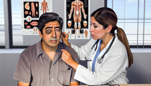
Middle-aged Hispanic patient with asymmetric facial weakness during a medical consultation. The Hispanic doctor is carefully examining the patient, focusing on the differential diagnosis of Bell's palsy versus stroke. The clinical setting features anatomical diagrams and medical tools, highlighting the importance of a thorough neurological examination of cranial nerves.

Bell's Palsy vs. Stroke: Essential Neurological Examination for Facial Weakness

Distinguishing between Bell's palsy and stroke is crucial for appropriate and timely treatment.

Read more
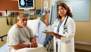
A middle-aged Hispanic patient sits on a hospital bed in the emergency room, displaying a slight look of confusion. A Hispanic female doctor, wearing a white coat and stethoscope, calmly explains while pointing to a brain diagram on a clipboard. The environment conveys urgency and care, emphasizing the critical importance of rapid assessment in stroke and TIA cases to prevent neurological deficits and facilitate timely thrombolysis.

Stroke vs. TIA: Urgent Assessment to Prevent Neurological Deficits and Optimize Thrombolysis Timing

Urgent assessment of stroke and TIA is crucial to prevent permanent neurological deficits.

Read more
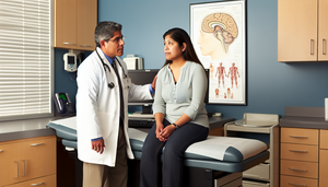
A middle-aged Hispanic doctor in a white coat and stethoscope attentively listens to a Hispanic female patient in her 30s during a medical consultation. The patient, with a worried expression, is seated on the examination table. On the wall, a diagram of the human brain highlights areas related to migraine and tension-type headache. The environment is professional and welcoming, ideal for a collaborative diagnostic approach focusing on headache management and photophobia.

Migraine vs. Tension-Type Headache: A Practical Guide for Diagnosis in Clinical Practice

Discover how to effectively differentiate between migraine and tension-type headache for optimal treatment.

Read more
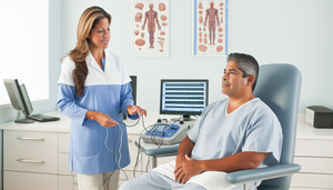
Middle-aged Hispanic patient in a neurology clinic, listening to a neurologist explaining the electromyography (EMG) procedure for ALS diagnosis. The EMG screen displays waveforms, highlighting a professional and collaborative approach to diagnosing progressive muscle weakness involving upper and lower motor neurons.

ALS Diagnosis: EMG and Clinical Criteria for Progressive Muscle Weakness in Upper and Lower Motor Neurons

Electromyography and Gold Coast criteria enhance the early diagnosis of ALS.

Read more
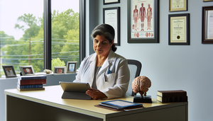
Middle-aged Hispanic female doctor in a medical office reviewing a patient file on a tablet, surrounded by medical books and a brain model on the desk, symbolizing neurological study. In the background, diplomas on the wall and a poster of the nervous system, with natural light streaming through a window, highlighting her expertise in multiple sclerosis treatment, including beta interferon, fingolimod, corticosteroids, and neurological rehabilitation.

Multiple Sclerosis Treatment: Disease Modifiers, Beta Interferon, Fingolimod, Corticosteroids, and Neurological Rehabilitation

Discover how disease-modifying treatments and rehabilitation enhance quality of life in multiple sclerosis.

Read more
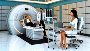
Modern medical office featuring a Hispanic physician in his 40s reviewing brain MRI scans with gadolinium on a screen, alongside a Hispanic patient in her 30s. Nearby, medical tools and a folder labeled "Evoked Potentials" are visible. This image emphasizes advanced technology and expertise in multiple sclerosis diagnosis, including demyelination assessment and CSF analysis for oligoclonal bands during relapses.

Diagnosing Multiple Sclerosis: MRI with Gadolinium, Oligoclonal Bands, and Relapses

The combination of brain MRI with gadolinium and evoked potentials enhances the diagnosis of multiple sclerosis.

Read more

Middle-aged Hispanic woman sitting at a desk, gently massaging her temples with a look of mild discomfort, illustrating symptoms of tension-type headache. The presence of a laptop and a glass of water on the desk suggests a work environment, highlighting the impact of stress management on bilateral headache and the importance of migraine differentiation in diagnosis.

Diagnosis of Tension-Type Headache: Clinical Features, Trigger Factors, and Migraine Differentiation

Explore how to differentiate tension-type headaches from migraines for effective stress management.

Read more
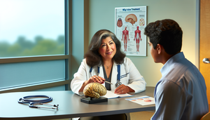
A medical consultation focused on migraine treatment, featuring a Hispanic doctor explaining options such as triptans and preventive beta-blockers to a Hispanic patient. The doctor holds a brain model, highlighting areas related to migraines. On the desk, there are brochures about "Triptans" and "Migraine Prevention" in Spanish. The setting is professional and welcoming, emphasizing a supportive approach to aura management and trigger control in migraine care.

Migraine Treatment: Triptans, Preventive Beta-Blockers, and Aura Management Strategies

Explore the use of triptans and preventive strategies for effective migraine management.

Read more
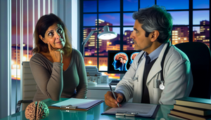
Middle-aged Hispanic woman in a modern medical consultation discussing migraine symptoms with a Hispanic doctor. The doctor listens attentively while holding a clipboard. In the office, a brain diagram on the computer highlights areas related to migraine diagnosis, pulsatile headache, and triggers. The professional and empathetic environment suggests collaboration in the neurological examination and management of aura symptoms.

Migraine Diagnosis: IHS Criteria, Differential Diagnosis, and Key Triggers for Pulsatile Headaches

Explore how IHS criteria and differential diagnosis enhance accuracy in migraine management.

Read more
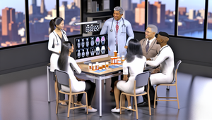
Multidisciplinary healthcare team collaborating in a modern medical office. A Hispanic neurologist discusses Parkinson's treatment options, including levodopa and dopamine agonists, with a pharmacist, a physical therapist, and a nurse. The table features medical charts, a laptop displaying brain scans, and labeled medication bottles for Parkinson's. This image emphasizes teamwork in the management of Parkinson's disease through neurorehabilitation and physical therapy.

Comprehensive Parkinson's Treatment: Pharmacological Options, Levodopa, Dopamine Agonists, and Multidisciplinary Management

Discover how the combination of levodopa, dopamine agonists, and physical therapy optimizes the management of Parkinson's disease.

Read more
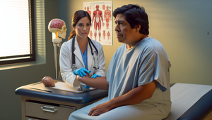
Middle-aged Hispanic patient in a medical consultation, sitting on an examination table with a thoughtful expression. He is wearing a light blue hospital gown. A Caucasian female doctor, dressed in a white coat and stethoscope, assesses the patient's resting tremor and bradykinesia. The setting is a medical office featuring charts and a human brain model, emphasizing the importance of accurate Parkinson's diagnosis and ruling out secondary parkinsonisms.

Parkinson's Diagnosis: Key Motor Signs, Differential Diagnosis, and Levodopa Response

Explore how to identify key motor signs and differentiate Parkinson's disease from other parkinsonisms.

Read more
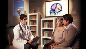
Image of a compassionate and professional medical environment focused on Alzheimer's treatment. A middle-aged Hispanic physician in a white coat and stethoscope explains a treatment plan to an elderly Hispanic female patient and her family, using a tablet that displays a brain diagram. The patient, a Hispanic woman in her 70s, appears engaged and hopeful. Her family, a young man and woman, listen attentively. The setting is warm and supportive, emphasizing a holistic and empathetic approach to Alzheimer's care, including cholinesterase inhibitors, memantine, and caregiver support in neuropsychiatry.

Alzheimer's Treatment: Cholinesterase Inhibitors, Memantine, and Comprehensive Caregiver Support in Neuropsychiatry

Discover how pharmacological treatments and a holistic approach enhance the quality of life in Alzheimer's patients.

Read more
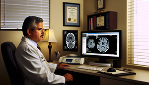
A middle-aged Hispanic neurologist carefully examines a magnetic resonance imaging scan of a brain on a computer screen, highlighting areas associated with Alzheimer's disease. In the background, a cognitive assessment test, including the MMSE, is visible on a desk. The scene conveys professionalism and dedication in Alzheimer's diagnosis, emphasizing the importance of memory tests and advanced imaging techniques like amyloid PET.

Alzheimer's Diagnosis: Cognitive Assessment with MMSE and Neuroimaging Techniques

The combination of memory tests and neuroimaging enhances the early diagnosis of Alzheimer's disease.

Read more