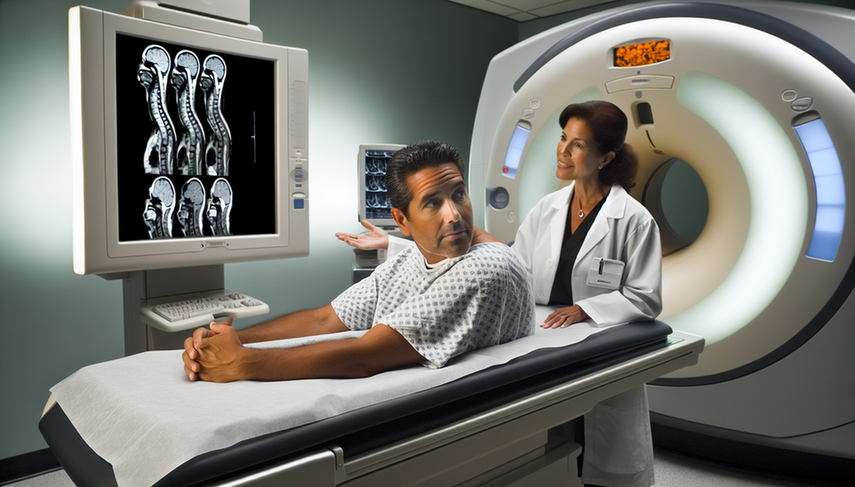
Diagnosis of Disc Herniation: MRI Findings and Radiating Low Back Pain from Nerve Compression

The disc herniation is a common cause of radiating low back pain and sciatica, significantly affecting patients' quality of life. Accurate diagnosis is crucial for determining the appropriate treatment, and magnetic resonance imaging (MRI) has established itself as the tool of choice for visualizing nerve compression and other structural alterations in the lumbar spine.
Diving Deeper into the Diagnosis
The lumbar MRI is essential for confirming the presence of a disc herniation and assessing its impact on adjacent nerve structures. Studies have shown that MRI not only allows for the identification of the herniation but also classifies it according to its severity, which is fundamental for deciding clinical management. For example, the Pfirrmann classification is a useful tool for evaluating disc degeneration and its correlation with clinical symptoms [1].
In addition to MRI, physical tests such as the Lasègue test and the Slump test are complementary methods that help evaluate the presence of radicular signs. A comparative study has shown that the Slump test has greater sensitivity in detecting disc herniations than the Lasègue test, suggesting its utility in clinical diagnosis [2].
The management of disc herniation can vary from conservative treatments to surgical interventions. The decision depends on the severity of symptoms and the response to initial treatment. In some cases, spontaneous regression of the disc herniation has been documented, reinforcing the importance of an initial conservative approach in many patients [3].
Conclusions
The diagnosis of lumbar disc herniation requires a combination of clinical evaluation and advanced imaging techniques such as MRI. The precise identification of nerve compression and its correlation with radicular signs are essential for guiding treatment. MRI not only provides detailed visualization of lumbar anatomy but also helps monitor changes in the disc and nerve roots over time [4]. The integration of physical tests and imaging allows for a more comprehensive and effective diagnostic approach.
Referencias
- [1] Lumbar Disc Herniation, the Association Between Quantitative Sensorial Test and Magnetic Resonance Imaging Findings
- [2] The sensitivity and specificity of the Slump and the Straight Leg Raising tests in patients with lumbar disc herniation
- [3] Spontaneous regression of a large sequestered lumbar disc herniation: a case report and literature review
- [4] Lumbar herniated disks
Created 6/1/2025
