Shingles vs. Contact Dermatitis: A Guide to Vesicular Lesions and Neuropathic Pain Management
Vesicular lesions on the skin can pose a diagnostic challenge, particularly when differentiating between shingles and contact dermatitis. Both conditions can present with vesicles, but their causes, clinical manifestations, and treatments are distinct. This article aims to provide a clear guide to distinguish between these two conditions, assisting physicians in making accurate diagnoses and selecting appropriate treatments.
Diving into Differential Diagnosis
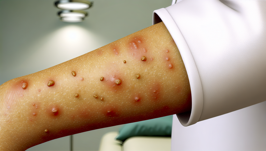
Shingles is a reactivation of the varicella-zoster virus, typically presenting as a painful, vesicular rash in a specific dermatome. Neuropathic pain is a common feature, and the vesicles often cluster on an erythematous base. A clinical case of shingles in the trigeminal nerve division illustrates the complexity of the differential diagnosis, as it can be confused with other vesiculobullous diseases of the mouth [1].
On the other hand, contact dermatitis is an inflammatory skin reaction caused by exposure to irritants or allergens. The lesions are usually more dispersed and may include vesicles on an erythematous base, but without the characteristic dermatomal pattern of shingles. A study on the infestation by Paederus species in Turkey highlights how these lesions can mimic both shingles and contact dermatitis, emphasizing the importance of a detailed clinical history for diagnosis [2].
Furthermore, it has been documented that the varicella-zoster virus can manifest in areas of pre-existing dermatitis, further complicating the diagnosis. A study on occult varicella shows how vesicular lesions can appear in sites of pre-existing dermatitis, suggesting the need to consider a viral infection in such cases [3].
Conclusions
Differentiating between shingles and contact dermatitis is crucial for the appropriate management of vesicular lesions. While shingles requires antiviral treatment and management of neuropathic pain, contact dermatitis is primarily managed by avoiding exposure to irritants and using topical corticosteroids. A careful clinical evaluation, supported by the patient's history and, when necessary, laboratory tests, is essential for accurate diagnosis.
Referencias
- [1] Herpes Zoster of the Third Division of the Trigeminal Nerve. A Clinical Pathologic Conference
- [2] An epidemicity of Paederus species in Cukurova region
- [3] Occult varicella
Created 6/1/2025