Melanoma vs. Seborrheic Keratosis: Key Insights for Early Diagnosis of Skin Cancer and Malignant Signs in Pigmented Lesions
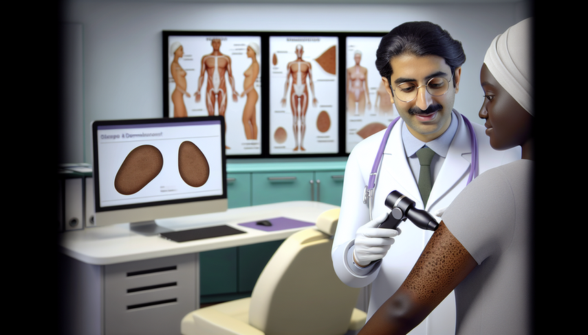
The early diagnosis of skin cancer is crucial for improving prognosis and survival rates among patients. Among the most common skin lesions that can be confused are melanoma and seborrheic keratosis. Although both may present pigmented lesions, their clinical and dermatoscopic characteristics are distinct. This article explores the key insights for differentiating these two conditions, emphasizing the importance of dermatoscopy and identifying signs of malignancy in the diagnostic process.

Dermatoscopic Features and Differential Diagnosis
Dermatoscopy is an invaluable tool for the differential diagnosis of pigmented skin lesions. In the case of melanoma, dermatoscopic signs include irregular dots and globules, asymmetrical pigmented follicular openings, and gray/black structures in a rhomboid shape. These findings are less common in seborrheic keratosis, which typically presents well-defined borders, superficial white or yellow scales, and cerebriform structures [1].
A recent study highlighted the utility of dermatoscopy in the differentiation of pigmented lesions on the face, underscoring the importance of identifying specific patterns for the accurate diagnosis of melanoma and other lesions [2]. Additionally, artificial intelligence and deep learning algorithms are emerging as promising tools to enhance diagnostic accuracy in skin lesions [3].
Conclusions and Recommendations
The early diagnosis of melanoma versus seborrheic keratosis is essential for appropriate patient management. Dermatoscopy remains a fundamental technique for distinguishing between these conditions, allowing for the identification of signs of malignancy that can guide clinical decision-making. The incorporation of new technologies, such as deep feature fusion and the use of neural networks, may further improve diagnostic accuracy and reduce unnecessary excision rates [4][5].
Referencias
- [1] Pigmented Macules on the Head and Neck: A Systematic Review of Dermoscopy Features
- [2] The utility and reliability of a deep learning algorithm as a diagnosis support tool in head & neck non-melanoma skin malignancies
- [3] Fusing fine-tuned deep features for skin lesion classification
- [4] Two-Stage Deep Neural Network via Ensemble Learning for Melanoma Classification
- [5] Reflectance confocal microscopy in the diagnosis of pigmented macules of the face: differential diagnosis and margin definition
Created 6/1/2025
