Middle-aged Hispanic patient in a modern medical consultation, attentively listening to a Hispanic doctor explaining the diagnosis of Fabry disease using a digital tablet. This interaction emphasizes effective communication in diagnosing Fabry disease, differentiating it from neurological conditions such as multiple sclerosis, while also addressing issues like neuropathic pain and corneal opacity related to alpha-galactosidase deficiency.

Fabry Disease: Diagnostic Keys and Differentiation of Neurological Processes in Alpha-Galactosidase Deficiency and Multiple Sclerosis

Discover how to differentiate Fabry disease from multiple sclerosis using essential diagnostic clues.

Read more
Middle-aged Hispanic patient in a modern medical examination room, discussing with a Hispanic doctor who holds a tablet displaying a diagram of the human body. An anatomical poster of the circulatory system is visible on the wall. This scene emphasizes the significance of a comprehensive evaluation in systemic amyloidosis, including cardiac assessment and abdominal fat biopsy for abnormal proteins, crucial for differentiating multiple myeloma.

Systemic Amyloidosis: Evaluating Multisystem Symptoms and Differentiating from Multiple Myeloma

Discover how to differentiate systemic amyloidosis from multiple myeloma through multisystem evaluation and advanced techniques.

Read more
Close-up of an 8-year-old Hispanic boy in a medical examination room, displaying cutaneous signs of tuberous sclerosis, such as hypomelanotic macules on the arms and face. This educational and professional image highlights skin features relevant for the differentiation of neurofibromatosis and emphasizes the importance of genetic evaluation in diagnosing conditions like seizures and facial angiofibromas.

Tuberous Sclerosis: Identifying Facial Angiofibromas and Differentiating from Neurofibromatosis in Seizure Patients

Discover how to identify cutaneous signs of tuberous sclerosis and differentiate them from other phakomatoses.

Read more
A middle-aged Hispanic man in a modern medical office, looking thoughtful, facing a Hispanic female doctor who is listening attentively. On the desk, a screen displays a brain scan image, indicating a discussion about Creutzfeldt-Jakob disease, rapidly progressive dementia, and the differentiation of encephalopathies, including the role of the 14-3-3 protein and EEG findings.

Creutzfeldt-Jakob Disease: Recognizing Rapidly Progressive Dementia and Differentiating from Other Encephalopathies Using 14-3-3 Protein and EEG

Discover how to differentiate Creutzfeldt-Jakob disease from other treatable encephalopathies using key markers.

Read more
Middle-aged Hispanic patient in a modern medical examination room, attentively listening to a Hispanic doctor who is explaining with a reassuring smile. The image conveys a professional and warm environment, ideal for discussing topics such as Shy-Drager syndrome, autonomic dysfunction, and atypical parkinsonism, with an emphasis on the importance of magnetic resonance imaging in differential diagnosis.

Shy-Drager Syndrome: Assessing Autonomic Dysfunction and Differential Diagnosis of Atypical Parkinsonism Using Magnetic Resonance Imaging

Discover how MRI and autonomic tests differentiate Shy-Drager syndrome from other parkinsonisms.

Read more
A middle-aged Hispanic patient with an attentive and slightly worried expression is seated on an examination table in a medical office. A Hispanic female doctor, wearing a white coat and stethoscope, is reassuringly explaining something while holding a clipboard. Behind them, there is an anatomical chart of the human nervous system. The scene reflects a professional and caring environment, pertinent to the discussion on Guillain-Barré syndrome, ascending weakness, EMG findings, and the differentiation from chronic polyradiculoneuropathy, including lumbar puncture considerations.

Early Detection of Guillain-Barré Syndrome: Differentiating from Chronic Polyradiculoneuropathy Using EMG and Lumbar Puncture

Early detection and differentiation between GBS and CIDP is crucial for optimal treatment.

Read more
A middle-aged Hispanic woman in an ophthalmologist's office, displaying concern as the Hispanic doctor explains something while pointing to an anatomical chart of the eye highlighting the optic nerve. On the desk, there are medical tools and a computer with a 3D model of the eye. The scene reflects a professional and modern environment, relevant for discussions on optic neuritis, sudden vision loss, and multiple sclerosis differentiation, including the evoked potential test.

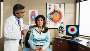
Optic Neuritis: Key Symptoms, Risk Assessment for Multiple Sclerosis, and the Role of Evoked Potential Tests in Sudden Vision Loss and Eye Pain

Early identification of optic neuritis is crucial for assessing the risk of multiple sclerosis.

Read more
A middle-aged Hispanic patient in a modern medical examination room, attentively listening to a Hispanic doctor explaining multisystem atrophy, dysautonomia, and the differentiation of parkinsonism from other parkinson plus syndromes using magnetic resonance imaging.

Multisystem Atrophy: Recognizing and Differentiating It from Other Parkinson Plus Syndromes Using MRI and Dysautonomia Insights

Discover how to differentiate multiple system atrophy from other Parkinson-plus syndromes using advanced techniques.

Read more
Brightly lit medical office where a middle-aged Hispanic doctor carefully examines a brain PET scan on a lightbox, highlighting areas associated with frontotemporal dementia. In the background, an older Hispanic woman observes the doctor. The scene reflects a professional and welcoming environment, emphasizing the importance of early diagnosis and differentiation from Alzheimer's disease, particularly in cases involving behavioral changes and progressive aphasia.

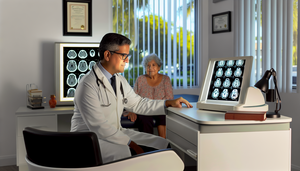
Frontotemporal Dementia: Early Diagnosis and Differentiation from Alzheimer's Disease through Behavioral Changes and Brain PET Imaging

Early differentiation between frontotemporal dementia and Alzheimer's enhances clinical management through neuroimaging and biomarkers.

Read more
A Hispanic male physician in his 40s, wearing a white coat and stethoscope, carefully examines a white matter MRI showing areas of demyelination, suggesting a diagnosis of Pelizaeus-Merzbacher. In the background, a Hispanic female physician in her 30s reviews genetic data, emphasizing the importance of early diagnosis and the differential diagnosis of leukodystrophies, including neonatal hypotonia and nystagmus.

Early Diagnosis of Pelizaeus-Merzbacher Disease: Differentiating from Other Leukodystrophies in Neonatal Hypotonia and Nystagmus with White Matter MRI

Early diagnosis of Pelizaeus-Merzbacher disease is crucial for differentiating it from other leukodystrophies and planning interventions.

Read more
A middle-aged Hispanic man in a modern medical office, focused on a medical chart. Next to him are medical books and a stethoscope, with anatomical charts on the wall, prominently featuring one of the brain. The scene reflects a medical research environment related to corticobasal degeneration, asymmetric rigidity, apraxia, and the differentiation of atypical parkinsonism through magnetic resonance imaging.

Corticobasal Degeneration: Recognizing Asymmetric Rigidity and Apraxia for Differentiation from Atypical Parkinsonism Using Magnetic Resonance Imaging

Discover how to differentiate corticobasal degeneration from other atypical parkinsonisms through clinical evaluation and magnetic resonance imaging.

Read more
Medical consultation in a modern hospital: a Hispanic physician in his 40s carefully reviews the medical history of a young Hispanic patient. On the desk, liver function test results and medical books are visible. An anatomical poster of the liver on the wall highlights the typical copper accumulation associated with Wilson's disease, emphasizing the importance of recognizing Kayser-Fleischer rings and ceruloplasmin levels in the differentiation of chronic hepatitis and neurological symptoms.

Early Diagnosis of Wilson's Disease: Differentiating from Chronic Hepatitis and Recognizing Neurological Symptoms

Early identification of Wilson's disease is crucial to prevent irreversible hepatic and neurological damage.

Read more
A middle-aged Hispanic patient sitting on an examination table in a modern medical office. A Caucasian doctor is carefully examining the patient's foot, illustrating a clinical evaluation of peripheral neuropathy. The wall features a diagram of the human nervous system, highlighting the importance of sensitivity testing and conduction velocity in the differentiation of diabetic polyneuropathy, with a focus on distal tingling.

Peripheral Neuropathy: Clinical Evaluation and Differentiation of Diabetic Polyneuropathy through Sensitivity Testing and Conduction Velocity Analysis

Discover how to differentiate diabetic polyneuropathy through effective clinical evaluation and sensitivity testing.

Read more
Medical consultation in a modern clinic: a Hispanic physician in his 40s examines the foot of a Hispanic teenager, pointing to a diagram of the nervous system on a tablet. On the wall, a poster about the anatomy of peripheral nerves. This scene emphasizes the importance of early diagnosis of Charcot-Marie-Tooth disease, hereditary peripheral neuropathy, and the role of electromyography in assessing foot deformities and differentiating ataxias.

Charcot-Marie-Tooth Disease: Early Diagnosis and Evaluation of Hereditary Peripheral Neuropathy through Electromyography and Differentiation of Ataxias

Early diagnosis of Charcot-Marie-Tooth disease enhances patient management and quality of life.

Read more
A middle-aged Hispanic patient with a concerned expression is sitting on an examination table while a Hispanic doctor, wearing a white coat and stethoscope, reassuringly explains something to him. She holds a clipboard, and on the wall, there is an anatomical chart of the nervous system, emphasizing the importance of early detection and differential diagnosis in neuromuscular disorders such as myasthenia gravis and Lambert-Eaton syndrome, including the role of the edrophonium test and anti-AChR antibodies in assessing fluctuating muscle weakness.

Myasthenia Gravis: Early Detection and Differentiation from Lambert-Eaton Syndrome Using Edrophonium Test and Anti-AChR Antibodies

Discover how to differentiate myasthenia gravis from Lambert-Eaton syndrome for accurate diagnosis.

Read more
A Hispanic girl approximately 10 years old is sitting in a consultation room with a middle-aged Hispanic female doctor, who is carefully examining the girl's hand. The scene reflects a professional and caring environment, relevant for the diagnosis of Sydenham's chorea, differentiation from other causes of involuntary movements, and the assessment of rheumatic fever through the ASO test.

Sydenham's Chorea: Diagnosis and Differentiation from Involuntary Movements and Huntington's Disease in Rheumatic Fever

Discover how to diagnose Sydenham's chorea and differentiate it from other causes of involuntary movements.

Read more
A middle-aged Hispanic man in an ophthalmologist's office, attentively observing a vision chart. The Hispanic female ophthalmologist points to the chart while explaining the significance of early detection and differentiation of retinal dystrophies, such as retinitis pigmentosa, which can lead to night blindness and tunnel vision.

Retinitis Pigmentosa: Early Symptom Identification and Differentiation from Other Retinal Dystrophies

Early detection of retinitis pigmentosa is crucial for accurate differential diagnosis.

Read more
An older Hispanic woman in an ophthalmology consultation, wearing glasses and attentively observing a vision chart. A Hispanic ophthalmologist explains something, pointing to the chart. The scene suggests an eye examination, crucial for the early diagnosis of age-related macular degeneration and differentiation of diabetic retinopathy, utilizing OCT and fluorescein angiography techniques.

Age-Related Macular Degeneration: Early Diagnosis and Differentiation from Diabetic Retinopathy Using OCT and Fluorescein Angiography

Early detection and advanced imaging techniques are crucial for differentiating AMD from DR.

Read more
Middle-aged Hispanic woman in a medical office, examining her swollen and reddened knuckles, indicative of rheumatoid arthritis. A smiling Hispanic physician explains the situation, while medical charts and a human hand model are visible on the desk, emphasizing the importance of differentiating between rheumatoid arthritis and osteoarthritis, including the role of rheumatoid factor and anti-CCP antibodies in the differential diagnosis.

Rheumatoid Arthritis: Evaluating Initial Symptoms and Differentiating from Osteoarthritis with Rheumatoid Factor and Anti-CCP Antibodies

Discover how to differentiate rheumatoid arthritis from osteoarthritis through symptoms, serological markers, and imaging.

Read more
Middle-aged Hispanic woman in a medical office, thoughtfully examining her hands with visible swelling in the knuckles, indicative of osteoarthritis. In the background, a Caucasian physician in a white coat holds a clipboard, attentively observing the patient in a modern and professional setting, highlighting the importance of X-ray evaluation for joint pain and rheumatoid arthritis differentiation amidst degenerative factors.

Osteoarthritis: Diagnostic Criteria and Differentiation from Rheumatoid Arthritis in Joint Pain Evaluation

Discover how to differentiate osteoarthritis from rheumatoid arthritis for more effective treatment.

Read more